J Korean Soc Radiol.
2013 Jan;68(1):17-20.
Incidental MRI Findings in Patients with Impaired Cognitive Function
- Affiliations
-
- 1Department of Radiology, College of Medicine, Inje University, Ilsan Paik Hospital, Goyang, Korea. hyj@paik.ac.kr
Abstract
- PURPOSE
This study aims to evaluate the incidental findings on brain MRI of patients with cognitive function impairments.
MATERIALS AND METHODS
We analyzed magnetic resonance (MR) findings of 236 patients with decreased cognitive function. MR protocols include conventional T2 weighted axial images, fluid attenuated inversion recovery axial images, T1 weighted coronal 3-dimensional magnetization-prepared rapid acquisition of gradient echo and diffusion tensor images. We retrospectively evaluated the signal changes that suggest acute/subacute infarction and space occupying lesions which show mass effect.
RESULTS
Incidental MR findings were seen in 16 patients. Nine patients (3.8%) showed increased signal intensity on trace map of diffusion tensor images suggesting acute/subacute infarctions. Space occupying lesions were detected in 7 patients, and 3 lesions (1.27%) had mass effect and edema and were considered clinically significant lesions that diminish cognitive functions.
CONCLUSION
Several incidental MR findings were detected in patients with decreased cognitive function, and the incidence of aucte/subacute infarctions were higher. Proper evaluations of MRI in patients with impaired cognitive functions will be helpful in early detection and management of ischemic lesions and space occupying lesions.
MeSH Terms
Figure
-
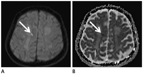
Fig. 1 A 78-year-old male with mild cognitive impairment. Focal increased signal intensity (A) is noted in right deep white matter on trace map of diffusion tensor images (arrow). Signal drop (arrow) is seen in ADC map (B). Note.-ADC = apparent diffusion coefficient
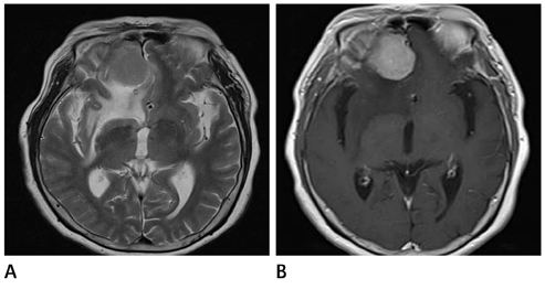
Fig. 2 A 70-year-old female with decreased cognitive function. Well defined mass lesion is seen in right frontal skull base on T2 weighted images (A) and strong contrast enhancement is noted on follow-up contrast enhanced T1 axial images (B). This lesion was surgically confirmed as meningioma.
Reference
-
1. Reitz C, Brayne C, Mayeux R. Epidemiology of Alzheimer disease. Nat Rev Neurol. 2011. 7:137–152.2. Dubois B, Feldman HH, Jacova C, Dekosky ST, Barberger-Gateau P, Cummings J, et al. Research criteria for the diagnosis of Alzheimer's disease: revising the NINCDS-ADRDA criteria. Lancet Neurol. 2007. 6:734–746.3. McKhann G, Drachman D, Folstein M, Katzman R, Price D, Stadlan EM. Clinical diagnosis of Alzheimer's disease: report of the NINCDS-ADRDA Work Group under the auspices of Department of Health and Human Services Task Force on Alzheimer's Disease. Neurology. 1984. 34:939–944.4. McKhann GM, Knopman DS, Chertkow H, Hyman BT, Jack CR Jr, Kawas CH, et al. The diagnosis of dementia due to Alzheimer's disease: recommendations from the National Institute on Aging-Alzheimer's Association workgroups on diagnostic guidelines for Alzheimer's disease. Alzheimers Dement. 2011. 7:263–269.5. Jack CR Jr, Dickson DW, Parisi JE, Xu YC, Cha RH, O'Brien PC, et al. Antemortem MRI findings correlate with hippocampal neuropathology in typical aging and dementia. Neurology. 2002. 58:750–757.6. Ikram MA, Vrooman HA, Vernooij MW, den Heijer T, Hofman A, Niessen WJ, et al. Brain tissue volumes in relation to cognitive function and risk of dementia. Neurobiol Aging. 2010. 31:378–386.7. Basser PJ, Mattiello J, LeBihan D. Estimation of the effective self-diffusion tensor from the NMR spin echo. J Magn Reson B. 1994. 103:247–254.8. Basser PJ, Mattiello J, LeBihan D. MR diffusion tensor spectroscopy and imaging. Biophys J. 1994. 66:259–267.9. Vermeer SE, Longstreth WT Jr, Koudstaal PJ. Silent brain infarcts: a systematic review. Lancet Neurol. 2007. 6:611–619.10. Matsui T, Nemoto M, Maruyama M, Yuzuriha T, Yao H, Tanji H, et al. Plasma homocysteine and risk of coexisting silent brain infarction in Alzheimer's disease. Neurodegener Dis. 2005. 2:299–304.11. Yamada K, Nagakane Y, Sasajima H, Nakagawa M, Mineura K, Masunami T, et al. Incidental acute infarcts identified on diffusion-weighted images: a university hospital-based study. AJNR Am J Neuroradiol. 2008. 29:937–940.12. Saini M, Ikram K, Hilal S, Qiu A, Venketasubramanian N, Chen C. Silent stroke: not listened to rather than silent. Stroke. 2012. 43:3102–3104.13. Katzman R. Katzman R, Rowe JW, editors. Diagnosis and management of dementia. Principles of geriatric neurology. 1992. Philadelphia: Davis;167–210.
- Full Text Links
-
- Actions
-
Cited
- CITED
-
- Close
- Share
-
- Similar articles
-
- Incidental Extramammary Findings on Preoperative Breast MRI in Breast Cancer Patients: A Pictorial Essay
- The Effectiveness of Brain MRI in Children and Adolescents with Headache
- Cognitive Rehabilitation in Traumatic Brain Injury
- Associations between Sleep and Work-Related Cognitive and Emotional Functioning in Police Employees
- Cognitive Dysfunction in non-hypoxemic COPD Patients
