Periodontal prosthesis on medically compromised patient with few remaining teeth: hybrid telescopic double crown with friction pin method
- Affiliations
-
- 1Department of Prosthodontics, School of Dentistry, Kyoungpook National University, Daegu, Republic of Korea. prosth95@knu.ac.kr
- KMID: 2321800
- DOI: http://doi.org/10.4047/jkap.2014.52.4.359
Abstract
- Successful results of treatments using double crown prostheses for the partially edentulous patients who have a few remaining teeth have been reported in several journals. A double crown removable partial denture can be an alternative treatment for the patients with a poor periodontal condition of remaining teeth. Since a double crown removable partial denture can be applied without the risk of surgical operation to the medically compromised patients with a poor periodontal condition which is inadequate for dental implants, it has psychological and economical advantages. In this case, there were sufficient remaining teeth to be restored with fixed prostheses in maxilla, while there were a few remaining teeth with a very poor periodontal condition so that it was almost impossible to restore with a clasp removable partial denture using these remaining teeth in mandible. In addition, the patient had the medical history of surgical operation due to osteomyelitis in the mandibular anterior areas a year ago, thus difficult to conduct an implant placement. The main objective of this report is to introduce our case because a double crown partial denture using a few mandibular remaining teeth showed satisfactory results in functional and esthetical aspects during more than two years follow-up period in this unfavorable condition.
MeSH Terms
Figure
-
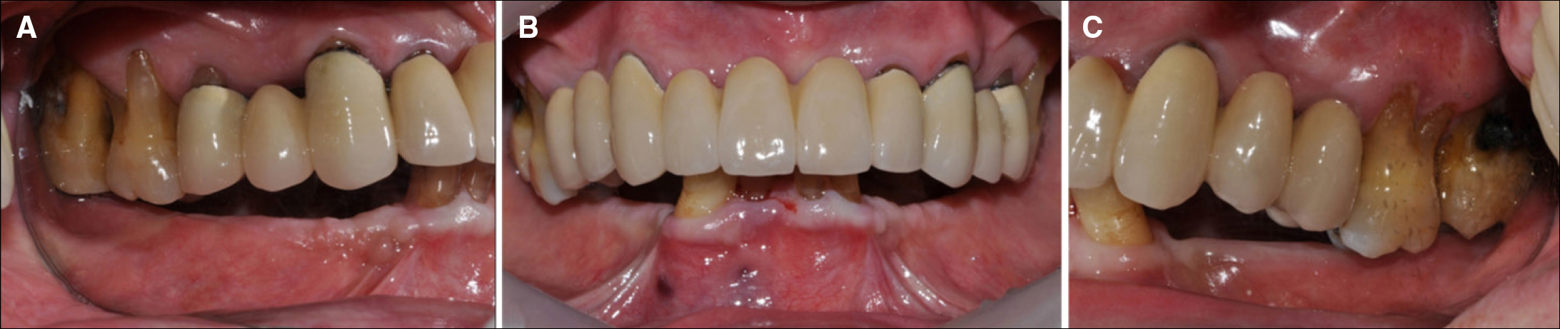
Fig. 1. Initial intraoral photograph. (A) Right buccal view, (B) Frontal view, (C) Left buccal view.

Fig. 2. Initial panoramic radiograph.
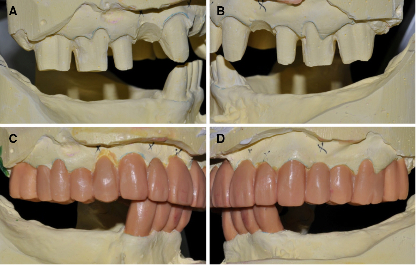
Fig. 3. The preparation of stone cast for provisional restoration and waxup procedure. (A, B) Right and left buccal view of the preparation of stone cast. (C, D) Right and left buccal view of the waxup procedure.

Fig. 4. Tooth preparation with waxup template. (A) Wax-up template sitting, (B) Maxillary tooth preparation, (C) Mandibular tooth preparation.
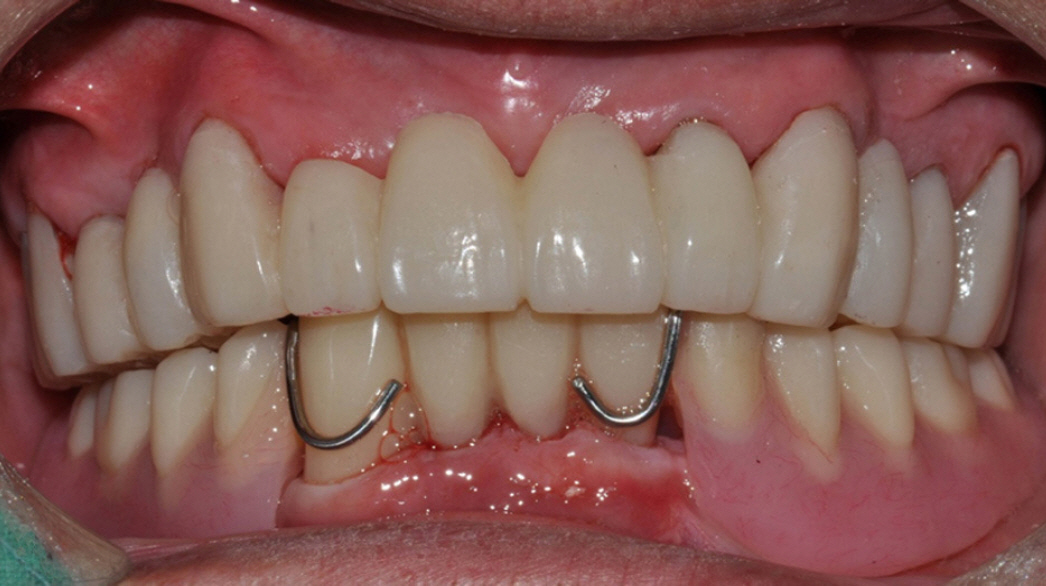
Fig. 5. Provisional restoration delivery.

Fig. 6. Inner crowns, Outer crown & Metal framework and Spark erosion procedure. (A) Inner crown, (B) Outer crown without major connector, (C) Spark erosion procedure. (D) Friction pin.

Fig. 7. Definitive restoration. (A) Right buccal view, (B) Frontal view, (C) Left buccal view.

Fig. 8. Final panoramic radiograph.
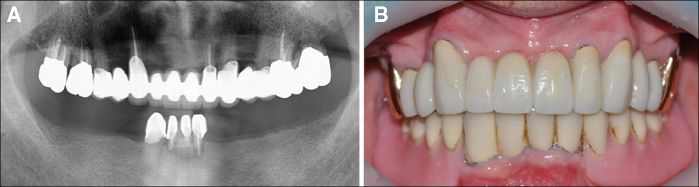
Fig. 9. Two-year follow-up panoramic radiograph & Intraoral photograph. (A) Panoramic radiograph, (B) Frontal view photograph.
Reference
-
1. Wenz HJ, Hertrampf K, Lehmann KM. Clinical longevity of removable partial dentures retained by telescopic crowns: outcome of the double crown with clearance fit. Int J Prosthodont. 2001; 14:207–13.2. Koller B, Att W, Strub JR. Survival rates of teeth, implants, and double crown-retained removable dental prostheses: a systematic literature review. Int J Prosthodont. 2011; 24:109–17.3. Saito M, Miura Y, Notani K, Kawasaki T. Stress distribution of abutments and base displacement with precision attachment-and telescopic crown-retained removable partial dentures. J Oral Rehabil. 2003; 30:482–7.4. Wenz HJ, Lehmann KM. A telescopic crown concept for the restoration of the partially edentulous arch: the Marburg double crown system. Int J Prosthodont. 1998; 11:541–50.5. Akagawa Y, Seo T, Ohkawa S, Tsuru H. A new telescopic crown system using a soldered horizontal pin for removable partial dentures. J Prosthet Dent. 1993; 69:228–31.
Article6. Weber H, Frank G. Spark erosion procedure: a method for extensive combined fixed and removable prosthodontic care. J Prosthet Dent. 1993; 69:222–7.
Article7. Murphy T. Compensatory mechanisms in facial height adjustment to functional tooth attrition. Aust Dent J. 1959; 4:312–23.
Article8. Murphy TR. The progressive reduction of tooth cusps as it occurs in natural attrition. Dent Pract Dent Rec. 1968; 19:8–14.9. Turner KA, Missirlian DM. Restoration of the extremely worn dentition. J Prosthet Dent. 1984; 52:467–74.
Article10. Hemmings KW, Howlett JA, Woodley NJ, Griffiths BM. Partial dentures for patients with advanced tooth wear. Dent Update. 1995; 22:52–9.11. Zarb G, Hobkirk J, Eckert S, Jacob R. Prosthodontic Treatment for Edentulous Patients. 13th ed.St. Louis: Mosby;2013. p. 180–203.12. Mauro Frandeani, Giancarlo Barducci. Esthetic rehabilitation in fixed prosthodontics. Volume 2. Honover Park: Quintessence Publishing Co.;2008. p. 154–15.13. Rivera-Morales WC, Mohl ND. Relationship of occlusal vertical dimension to the health of the masticatory system. J Prosthet Dent. 1991; 65:547–53.
Article14. Dahl BL, Krogstad O. Long-term observations of an increased occlusal face height obtained by a combined orthodontic/prosthetic approach. J Oral Rehabil. 1985; 12:173–6.
Article15. Wenz HJ, Lehmann KM. A telescopic crown concept for the restoration of the partially edentulous arch: the Marburg double crown system. Int J Prosthodont. 1998; 11:541–50.16. Heckmann SM, Schrott A, Graef F, Wichmann MG, Weber HP. Mandibular two-implant telescopic overdentures. Clin Oral Implants Res. 2004; 15:560–9.17. Zafiropoulos GG, Rebbe J, Thielen U, Deli G, Beaumont C, Hoffmann O. Zirconia removable telescopic dentures retained on teeth or implants for maxilla rehabilitation. Three-year observation of three cases. J Oral Implantol. 2010; 36:455–65.
Article18. Weber H, Frank G. Spark erosion procedure: a method for extensive combined fixed and removable prosthodontic care. J Prosthet Dent. 1993; 69:222–7.
Article
- Full Text Links
-
- Actions
-
Cited
- CITED
-
- Close
- Share
-
- Similar articles
-
- A clinical case of hybrid telescopic double crown using friction pin with an isolated few remaining teeth
- Clinical Report by using hybrid telescopic double crown Removable Partial Denture on a few remaining teeth with severe periodontal disease
- Prosthetic treatment for patient with anterior overbite and partial edentulism using maxillary hybrid telescopic double crown RPD and mandibular fixed prostheses: A 11-yr follow-up
- Treatment of the cleft lip and palate patient with few remaining posterior teeth using hybrid telescopic crown denture
- Causes of failures of long-term used double crown denture and new rehabilitation with dental implant and tooth combined denture using remaining teeth and implants
