Marginal and internal fitness of three-unit zirconia cores fabricated using several CAD/CAM systems
- Affiliations
-
- 1Department of Prosthodontics, School of Dentistry, Pusan National University, Yangsan, Korea.
- 2Institute for Clinical Dental Research, Korea University, Seoul, Korea. swshin@korea.ac.kr
Abstract
- PURPOSE
This study was aimed to compare the margin and internal fitness of 3-unit zirconia bridge cores fabricated by several CAD/CAM systems using replica technique.
MATERIALS AND METHODS
Three unit-bridge models in which upper canine and upper second premolar were used as abutments and upper first premolar was missed, were fabricated. Fourty models were classified into 4 groups (Cerasys(R) (Group C), Dentaim(R) (Group D), KaVo Everest(R) (Group K), Lava(TM)(Group L)), and zirconia cores were fabricated by each company. Sixteen points were measured on each abutment by replica technique. Statistical analysis was accomplished with two way ANOVA and Dunnett T3 (alpha=.05).
RESULTS
In most systems, there was a larger gap on inter margin than outer margin. In the Group K, overall fitness was excellent, but the incisal gap was very large. In the Group C, marginal gap was significantly larger than Group K, but overall internal gap was uniform (P<.05). The axial gap was under 100 microm in all system. The difference between internal and external gap was small on Group L and C. However, internal gap was significantly larger than external gap in Group D (P<.05). The fitness of canine was better than second premolar among abutments (P<.05).
CONCLUSION
The marginal and internal gap was within the clinically allowed range in all of the three systems. There was a larger gap on second premolar than canine on internal and marginal surface. In most systems, there was a larger gap on occlusal surface than axial surface.
Keyword
MeSH Terms
Figure
-
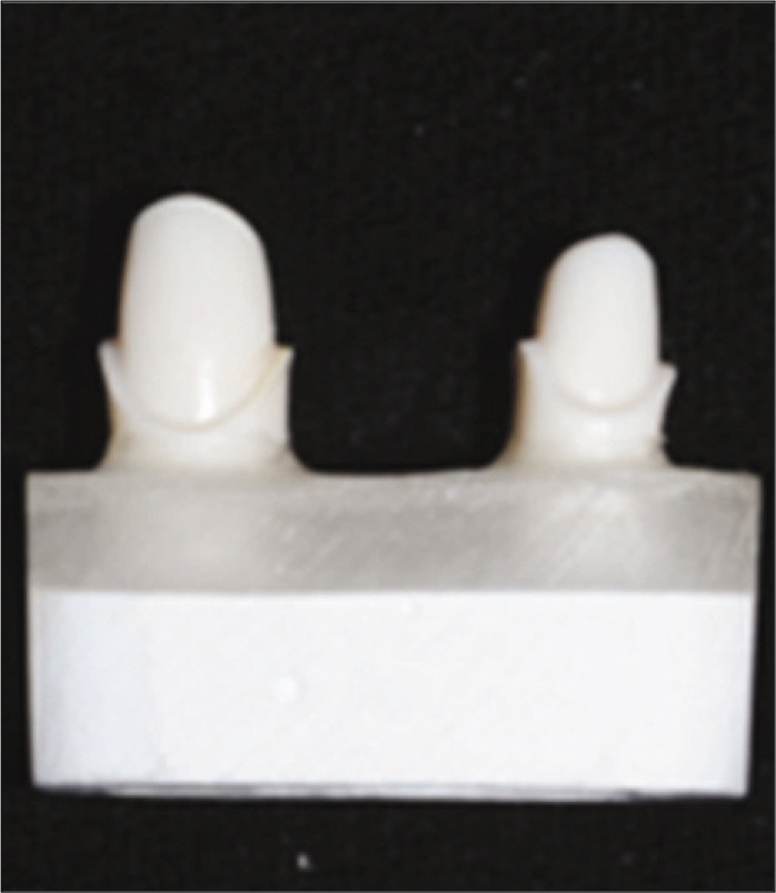
Fig. 1. Master model for replication with titanium block: Used dentiform teeth with 1.0 mm deep chamfer margin and 12 degree axial wall.
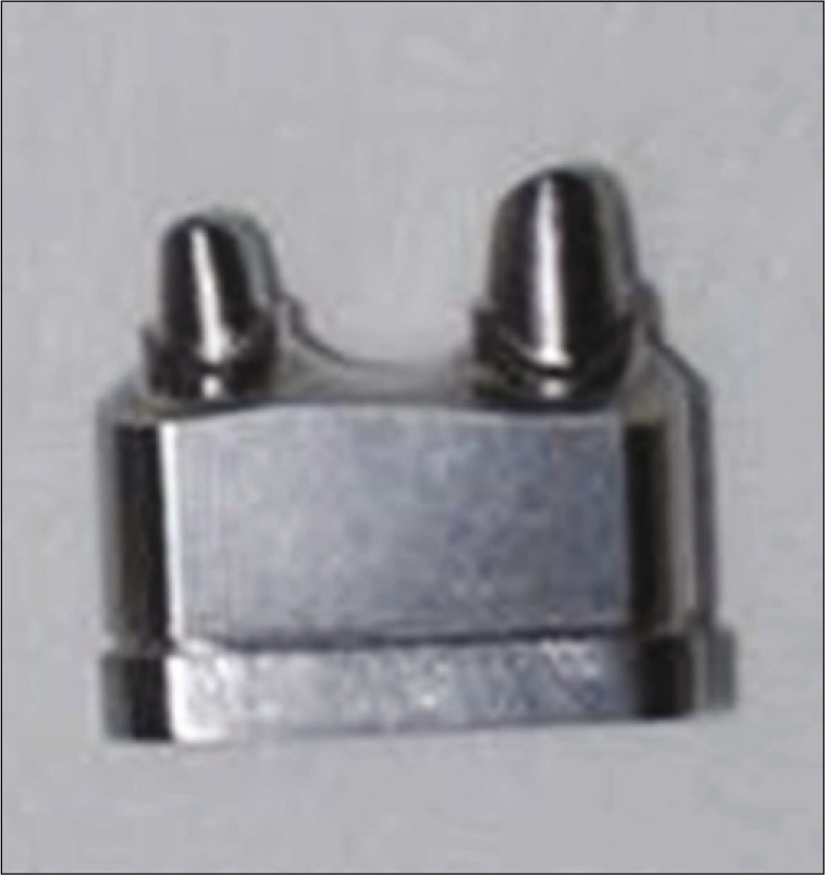
Fig. 2. Replica models with titanium block. This was fabricated with CAD/CAM system (Addtech Co., Seoul, Korea), and replicated master models.
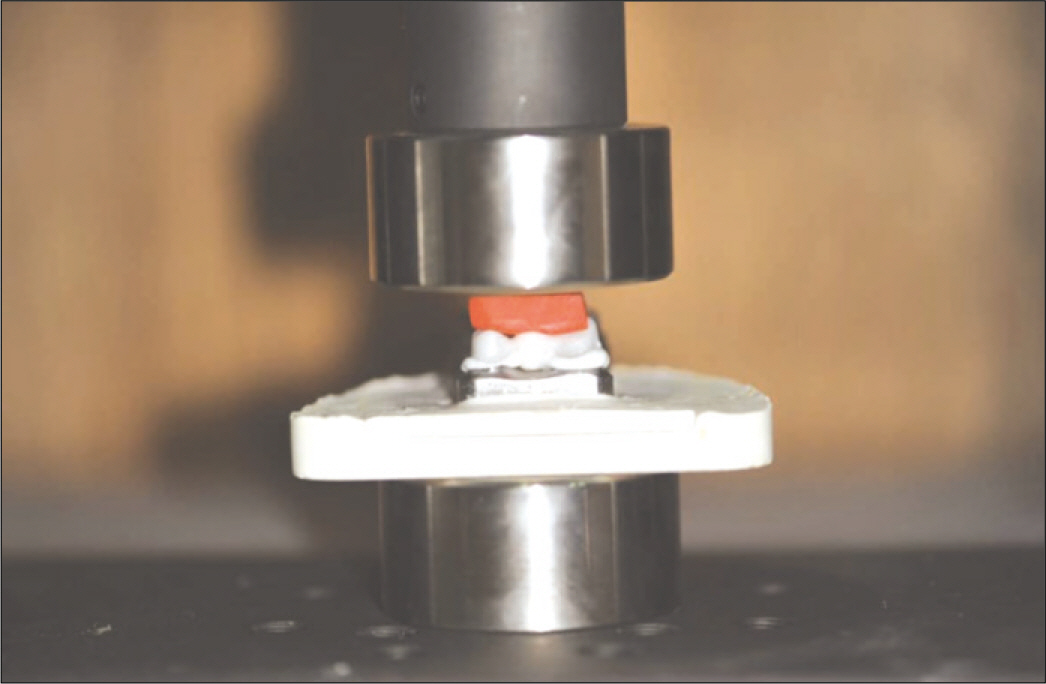
Fig. 3. The constant seating force (25 N) was maintained using a universal testing machine for 5 min.
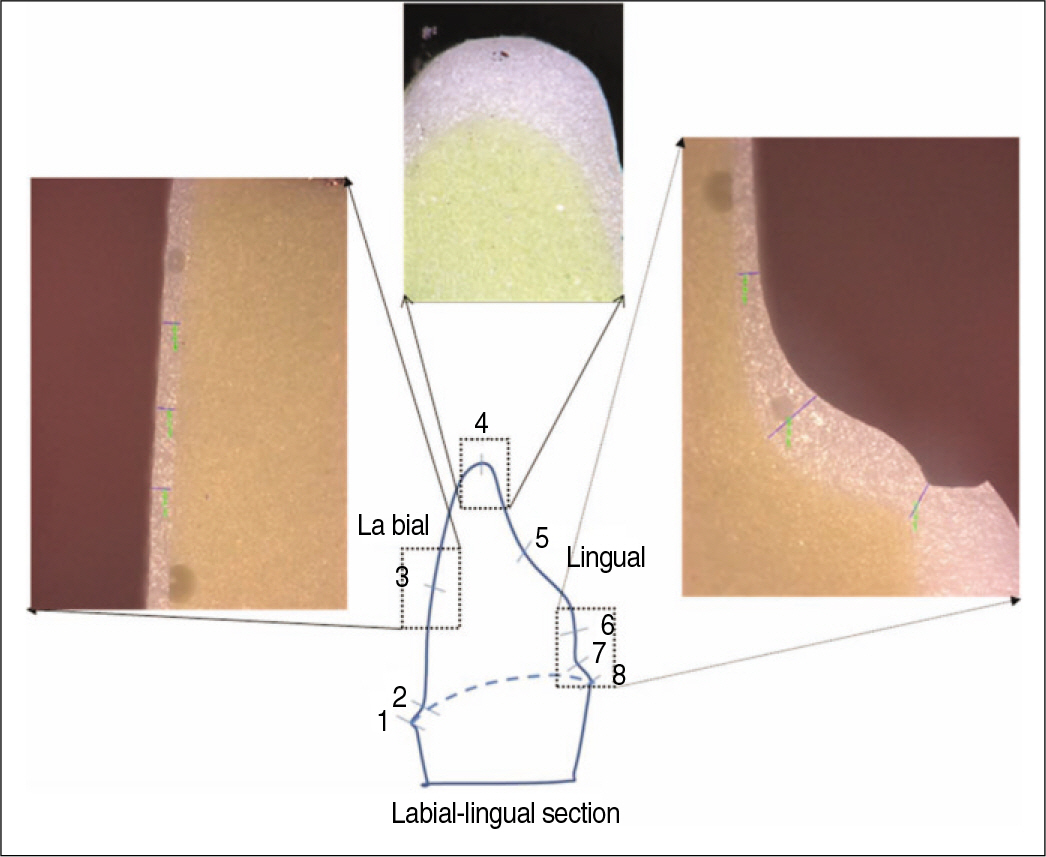
Fig. 4. Captured figures after sectioning with bucco-lingual direction using “Replica technique” .
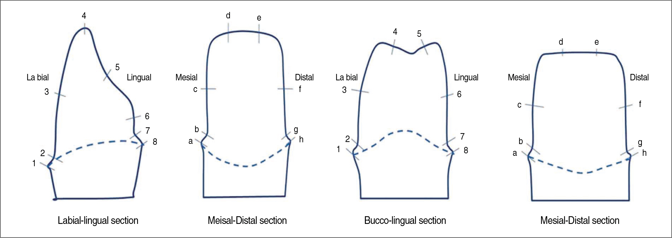
Fig. 5. Reference points to measure the thickness of fit checker.
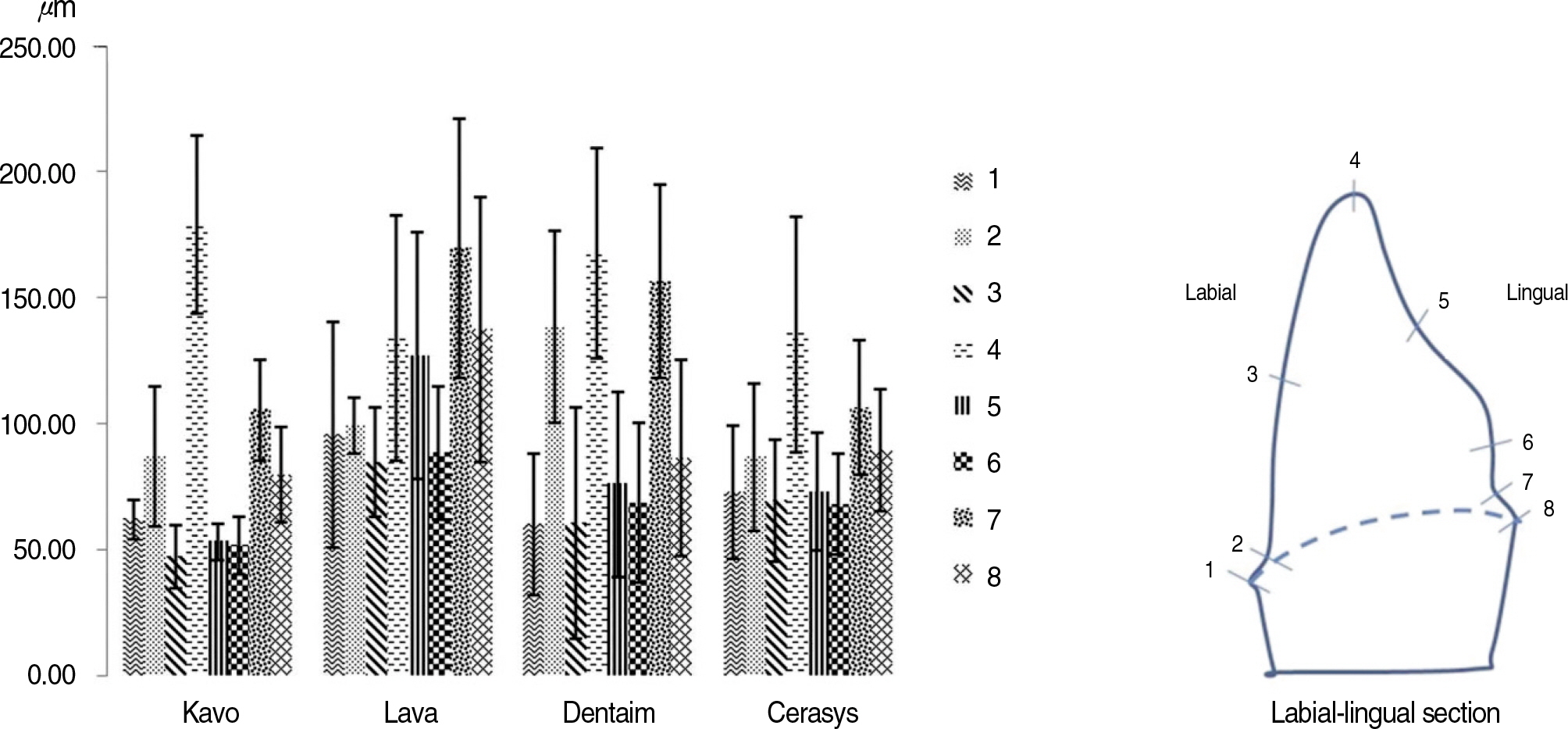
Fig. 6. Mean values and SD at each point in canine bucco-lingual section.
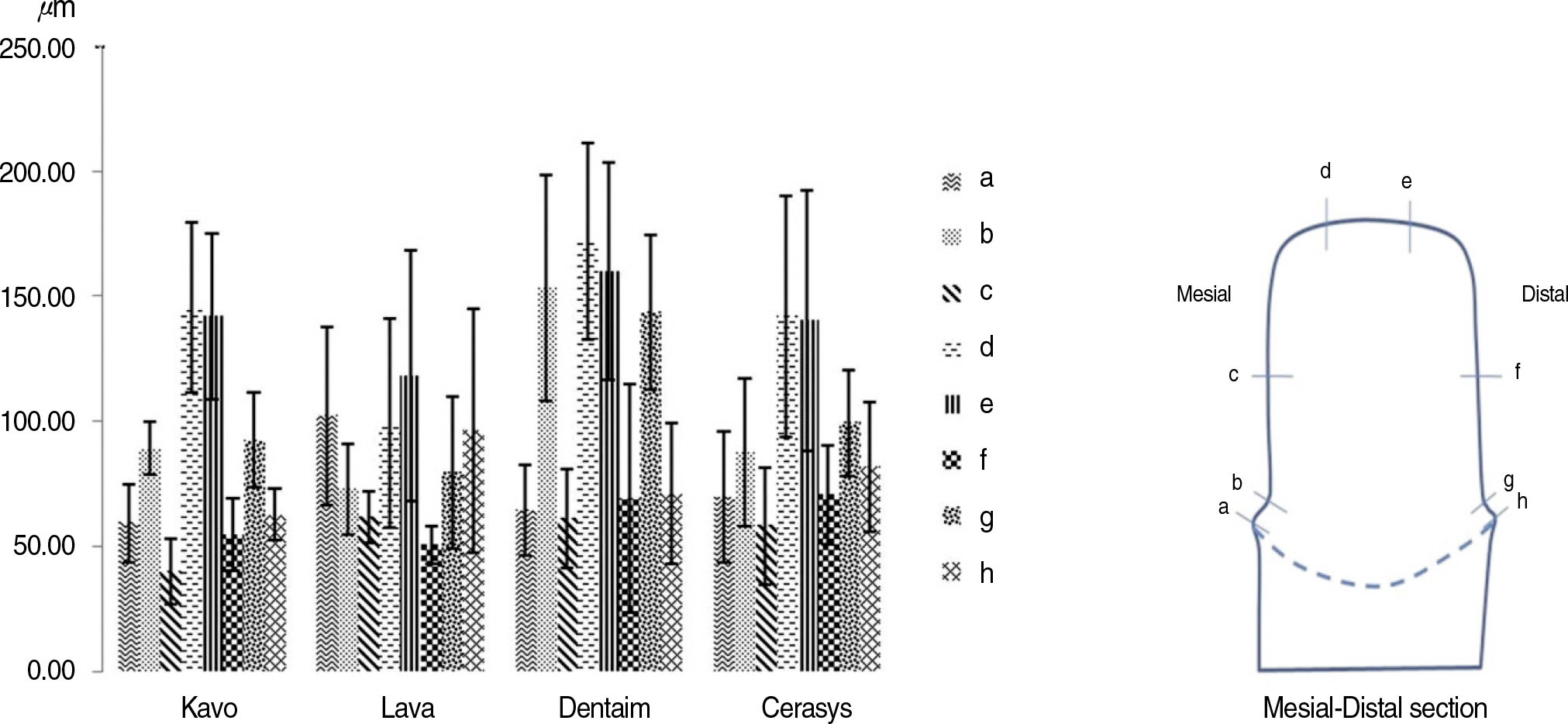
Fig. 7. Mean values and SD at each point in canine mesio-distal section.
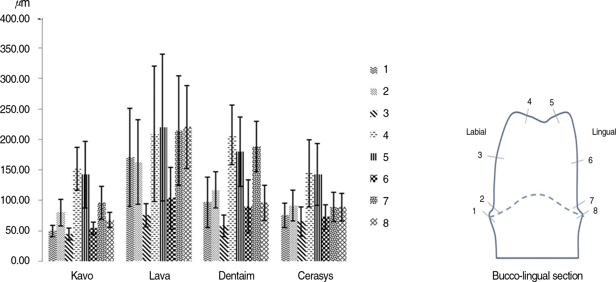
Fig. 8. Mean values and SD at each point in 2nd premolar bucco-lingual section.
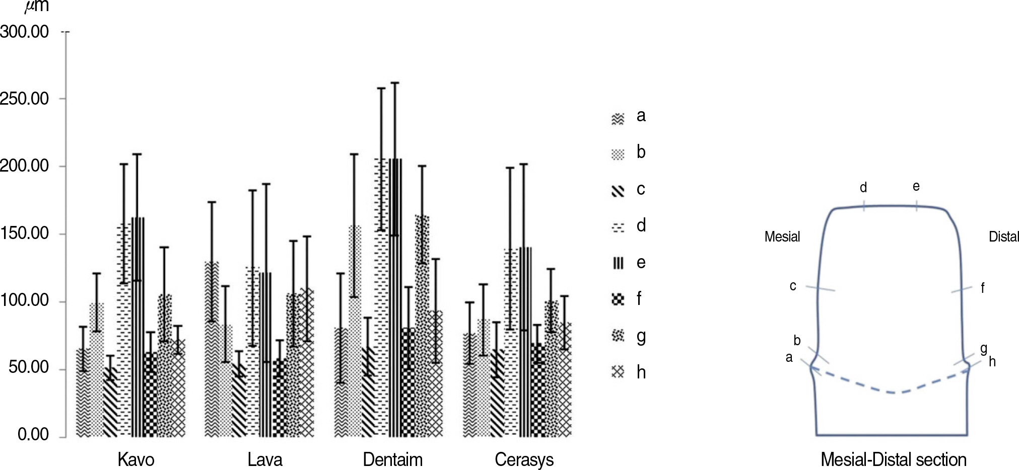
Fig. 9. Mean values and SD at each point in 2nd premolar mesio-distal section.
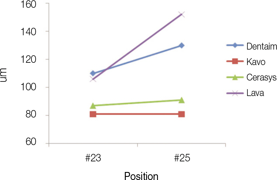
Fig. 10. Estimated means of marginal gap in each position and system.
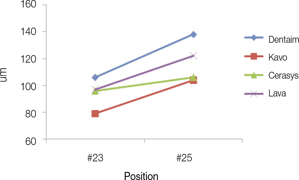
Fig. 11. Estimated means of internal gap in each position and system.
Reference
-
1.Bindle A., Mormann WH. Marginal and internal fit of allceramic CAD/CAM crown-coping on chamfer preparations. J Oral Rehabil. 2005. 32:441–7.2.Tinschert J., Natt G., Mautsch W., Spiekermann H., Anusavice KJ. Marginal fit of alumina-and zirconia-based fixed partial dentures produced by a CAD/CAM system. Oper Dent. 2001. 26:367–74.3.Yang JH., Yeo IS., Lee SH., Han JS., Lee JB. Marginal fit of celay/In-Ceram, Conventional In-Ceram and Empress 2 all-ceramic single crowns. J Korean Acad Prosthodont. 2002. 40:131–9.4.May KB., Russell MM., Razzoog ME., Lang BR. Precision of fit: the Procera AllCeram crown. J Prosthet Dent. 1998. 80:394–404.
Article5.Tinschert J., Natt G., Mautsch W., Spiekermann H., Anusavice KJ. Marginal fit of alumina-and zirconia-based fixed partial dentures produced by a CAD/CAM system. Oper Dent. 2001. 26:367–74.6.Hertlein G., Hoscheler S., Frank S., Suttor D. Marginal fit of CAD/CAM manufactured all ceramic prosthesis. J Dent Res. 2001. 80:42–4.7.Rekow ED. High-technology innovations-and limitations-for restorative dentistry. Dent Clin North Am. 1993. 37:513–24.8.Valderrama S., Van Roekel N., Andersson M., Goodacre CJ., Munoz CA. A comparison of the marginal and internal adaptation of titanium and gold-platinum-palladium metal ceramic crowns. Int J Prosthodont. 1995. 8:29–37.9.Sturdevant JR., Bayne SC., Heymann HO. Margin gap size of ceramic inlays using second-generation CAD/CAM equipment. J Esthet Dent. 1999. 11:206–14.
Article10.Gardner FM. Margins of complete crowns-literature review. J Prosthet Dent. 1982. 48:396–400.
Article11.Hung SH., Hung KS., Eick JD., Chappell RP. Marginal fit of porcelain-fused-to-metal and two types of ceramic crown. J Prosthet Dent. 1990. 63:26–31.
Article12.Wang CJ., Millstein PL., Nathanson D. Effects of cement, cement space, marginal design, seating aid materials, and seating force on crown cementation. J Prosthet Dent. 1992. 67:786–90.
Article13.Molin M., Karlsson S. The fit of gold inlays and three ceramic inlay systems. A clinical and in vitro study. Acta Odontol Scand. 1993. 51:201–6.
Article14.Sturdevant JR., Bayne SC., Heymann HO. Margin gap size of ceramic inlays using second-generation CAD/CAM equipment. J Esthet Dent. 1999. 11:206–14.
Article15.Huh JB., Park CG., Kim HY., Park CK., Shin SW. Evaluation using replica technique on the marginal and internal fitness of zirconia cores by several CAD/CAM systems. J Korean Acad Prosthodont. 2010. 48:135–42.
Article16.Carter JM., Sorensen SE., Johnson RR., Teitelbaum RL., Levine MS. Punch shear testing of extracted vital and endodontically treated teeth. J Biomech. 1983. 16:841–8.
Article17.Strawn SE., White JM., Marshall GW., Gee L., Goodis HE., Marshall SJ. Spectroscopic changes in human dentine exposed to various storage solutions-short term. J Dent. 1996. 24:417–23.18.Koo JY., Lim JH., Cho IH. Marginal fidelity according to the margin types of all ceramic crowns. J Korean Acad Prosthodont. 1997. 35:445–57.19.Pera P., Gilodi S., Bassi F., Carossa S. In vitro marginal adaptation of alumina porcelain ceramic crowns. J Prosthet Dent. 1994. 72:585–90.
Article20.Belser UC., MacEntee MI., Richter WA. Fit of three porcelain-fused-to-metal marginal designs in vivo: a scanning electron microscope study. J Prosthet Dent. 1985. 53:24–9.
Article21.Davis DR. Comparison of fit of two types of all-ceramic crowns. J Prosthet Dent. 1988. 59:12–6.
Article22.Abbate MF., Tjan AH., Fox WM. Comparison of the marginal fit of various ceramic crown systems. J Prosthet Dent. 1989. 61:527–31.
Article23.Wu JC., Wilson PR. Optimal cement space for resin luting cements. Int J Prosthodont. 1994. 7:209–15.24.Brukl CE., Nicholson JW., Norling BK. Crown retention and seating on natural teeth with a resin cement. J Prosthet Dent. 1985. 53:618–22.
Article25.Yu JH., Kim YC., Kang DW. A study on the marginal fidelities and fracture strength of IPS Empress 2 ceramic crowns. J Korean Acad Prosthodont. 2000. 38:606–17.26.Sorensen JA. A standardized method for determination of crown margin fidelity. J Prosthet Dent. 1990. 64:18–24.
Article27.Moon BH., Yang JH., Lee SH., Chung HY. A study on the marginal fit of all-ceramic crown using ccd camera. J Korean Acad Prosthodont. 1998. 36:273–92.28.Rahme HY., Tehini GE., Adib SM., Ardo AS., Rifai KT. In vitro evaluation of the "replica technique" in the measurement of the fit of Procera crowns. J Contemp Dent Pract. 2008. 9:25–32.
- Full Text Links
-
- Actions
-
Cited
- CITED
-
- Close
- Share
-
- Similar articles
-
- Comparative study in marginal adaptation of zirconia cores fabricated with 3 different CAD/CAM systems
- Evaluation using Replica Technique on the marginal and internal fitness of zirconia cores by several CAD/CAM systems
- The fit of zirconia core fabricated with CAD/CAM system
- Marginal fit of the digident CAD/CAM zirconia ceramic crowns
- Comparison of occusal aspects in monolithic zirconia crown before and after occlusal adjustment during intraoral try-in: a case report
