J Korean Acad Periodontol.
2009 Dec;39(4):425-429.
Treatment of periodontal lesion caused by palatogingival groove in maxillary lateral incisor: case reports
- Affiliations
-
- 1Department of Periodontology, School of Dentistry, Kyungpook National University, korea. leejm@knu.ac.kr
Abstract
- PURPOSE
The palatogingival groove is a developmental anomaly of the incisor teeth, which often presents severe localized periodontal disease. The purpose of this study was to evaluate the clinical outcome of palatogingival groove-associated periodontal lesion following flap operation with glass ionomer filling. METHODS: Four patients with periodontal lesion associated with the palatogingival groove were chosen for this case study. Clinical indices were taken and radiographic exam was performed at the baseline of the study and four patients were treated by flap operation with GI filling. Post-surgical visits were scheduled at regular intervals to check clinical and radiographic changes. RESULTS: Symptoms and signs of periodontal lesion were almost completely resolved with improvement of periodontal indices CONCLUSIONS: Flap operation with direct glass ionomer restoration is thought to be an acceptable method which can produce favorable results in the treatment of periodontal lesion caused by palatogingival groove on the maxillary lateral incisor.
Keyword
MeSH Terms
Figure
-
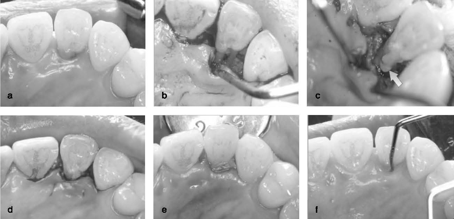
Figure 1 Clinical photograph of first patient. a: Preoperative, #22. b: Flap reflection & debridement. c: Glass ionomer filling. d: Suture. e: Postoperative, 1w eek. f: Postoperative, 11 months.

Figure 2 Clinical photograph of second patient. a: Preoperative, #12. b: Flap reflection & debridement. c: Glass ionomer filling. d: Suture. e: Postoperative, 1w eek. f: Postoperative, 4 months.

Figure 3 Clinical photograph of third patient. a: Initial. b: Flap reflection. c: #22 Debridement. d: Glass ionomer filling. e: Suture. f: Postoperative, 5 years.
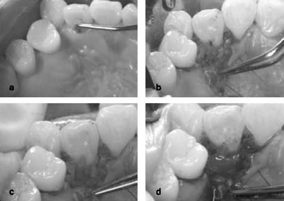
Figure 4 Clinical photograph of fourth patient. a: Initial, #12. b: Flap reflection. c: Debridement & groove enlargement. d: Emdogain application.
Reference
-
1. Ballal NV, Jothi V, Bhat KS, Bhat KM. Salvaging a tooth with a deep palatogingival groove: an endoperio treatment - a case report. International Endodontic Journal. 2007. 40:808–817.
Article2. Gound TG, Maze GI. Treatment options for the radicular lingual groove: A review and discussion. Pract Periodont Aesteht Dent. 1998. 10:369–375.3. Atkinson SR. The permanent maxillary lateral incisor. American J Orthodont. 1943. 29:685–688.
Article4. Everett FG, Kramer GM. The disto-lingual groove in the maxillar lateral incisor: A Periodontal hazard. J Periodontol. 1972. 43:352–361.
Article5. Withers JA, Brunsvold MA, Jilloy WJ, Rahe AJ. The relationship of palato-gingival grooves to localized periodontal disease. J Periodontol. 1981. 52:41–44.
Article6. Kogon SL. The prevalence, location and conformation of palato-radicular grooves in maxillary incisors. J Periodontol. 1986. 57:231–234.
Article7. Lara VS, Consolaro A, Bruce RS. Macroscopic and microscopic analysis of the palato-gingival groove. J Endodont. 2000. 26:345–350.
Article8. Meister F Jr, Keating K, Gerstein H, Mayer JC. Successful treatment of a radicular lingual groove: Case report. J Endodont. 1983. 9:561–564.9. Friedman S, Goultschin J. The radicular palatal groove- A therapeutic modality. Endodont Dent Traumatol. 1988. 4:282–286.10. Vermeersch G, Leloup G, Delme M, Vreven J. Antibacterial activity of glass-ionomer cements, compomers and resin composites: relationship between acidity and material setting phase. J Oral Rehabil. 2005. 32:368–374.
Article11. Vermeersch G, Leloup G, Vreven J. Fluoride release from glass-ionomer cements, compomers and resin composites. J Oral Rehabil. 2001. 28:26–32.
Article12. Dragoo MR. Resin-ionomer and hybrid-ionomer cements: Part I. Comparison of three materials for the treatment of subgingival root lesions. Int J Periodontics Restorative Dent. 1996. 16:594–601.13. Dragoo MR. Resin-ionomer and hybrid-ionomer cements: part II, human clinical and histologic wound healing responses in specific periodontal lesions. Int J Periodontics Restorative Dent. 1997. 17:75–87.14. Jeng HJ, Lu HKJ, Hou LT. Treatment of an osseous lesion associated with a severe palato-radicular groove: A case report. J Periodontol. 1992. 63:708–711.
Article15. Al-hezaimi K, Naghshbandi J, Simon JH, Rotstein I. Successful treatment of a radicular groove by intentional replantation and Emdogain therapy: four years follow-up. Oral Surg Oral Med Oral Patho Oral Radiol Endod. 2009. 107:e82–e85.
Article
- Full Text Links
-
- Actions
-
Cited
- CITED
-
- Close
- Share
-
- Similar articles
-
- Treatment of a lateral incisor anatomically complicated with palatogingival groove
- The palato-gingival groove - anatomical anomaly occurred in maxillary lateral incisors: case reports
- Recognition and management of palatogingival groove for tooth survival: a literature review
- Management of apicomarginal defect in esthetic region associated with a tooth with anomalies
- A study on the pattern of movement during retraction of maxillary central incisor by finite element method
