The palato-gingival groove - anatomical anomaly occurred in maxillary lateral incisors: case reports
- Affiliations
-
- 1Department of Conservative Dentistry, College of Dentistry, Wonkwang University, Korea. mksdd@wonkwang.ac.kr
Abstract
- This report describes clinical cases of a palato-gingival groove on a maxillary lateral incisor with associated localized periodontal disease and pulp necrosis. The tooth of the first case was extracted because of severe bone destruction. The palato-gingival groove of the second case was eliminated using a round bur, and the resulting defect was filled with synthetic graft and covered by an absorbable membrane. Both diagnosis and treatment of palato-gingival groove were very difficult and usually extraction of the involved tooth is the treatment of choice, but combined endodontic-periodontic treatment allowed the tooth to be saved.
Figure
-

Figure 1 Pretreatment images. (A) Photograph demonstrating the facial sinus tract. (B) Probing depth (8 mm) at the cingulum. (C) Periapical radiograph showing a gutta-percha cone tracing the facial sinus tract.
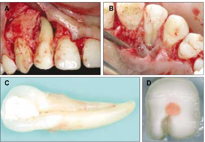
Figure 2 (A) and (B) The osseous defect associated with the palato-gingival groove (arrow). (C) Lingual side of extracted tooth shows palato-gingival groove running along entire length of root. (D) Cross sectional view of extracted tooth at 5 mm level from CEJ shows the communication between the root canal system and the groove.
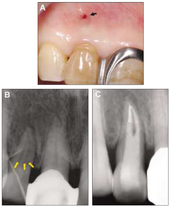
Figure 3 (A) Preoperative clinical view demonstrating the facial sinus tract (arrow) and discoloration of lateral incisor. (B) Periapical radiograph showing a gutta-percha cone tracing the facial sinus tract to the periradicular radiolucency associated with the right maxillary lateral incisor. Three vertically oriented radiolucent lines are also evident within lateral incisor (arrows). (C) Nonsurgical endodontic treatment; gutta-percha on apical one-third and resin core on middle and coronal one-third.
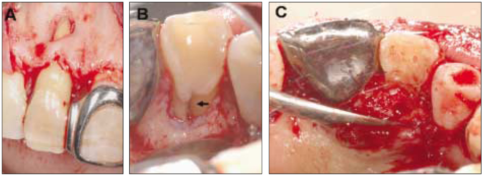
Figure 4 (A) The fenestration of the cortical plate on the facial aspect. (B) The palatal bony defect associated with the palatal groove (arrow). (C) The palatal groove after odontoblasty and filling of the bony defect with freeze-dried bone allograft.
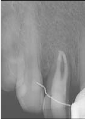
Figure 5 Periapical radiograph of tooth 6 months after treatment.
Reference
-
1. American Association of Endodontists. Glossary of endodontic terms. 2003. 7th ed. Chicago, IL:2. Lee KW, Lee EC, Poon RY. Palato-gingival grooves in maxillary incisors. A possible predisposing factor to localized periodontal disease. Br Dent J. 1968. 124:14–18.3. Simon JH, Glick DH, frank AL. Predictable endodontic and periodontic failure as a result of radicular anomalies. Oral Surg Oral Med Oral Pathol. 1971. 31:823–826.
Article4. Ennes JP, Lara VS. Comparative morphological analysis of the root developmental groove with the palate-gingivalgroove. Oral Dis. 2004. 10:378–382.
Article5. Everett FG, Kramer GM. The disto-lingual groove in the maxillary lateral incisor; a periodontal hazard. J Periodontol. 1972. 43:352–361.
Article6. August DS. The radicular lingual groove: an overlooked differential diagnosis. J Am Dent Assoc. 1978. 96:1037–1039.
Article7. Kogon SL. The prevalence, location and conformation of palato-radicular grooves in maxillary incisors. J Periodontol. 1986. 57:231–234.
Article8. Pecora JD, Sousa Neto MD, Santos TC, Saquy PC. In vitro study of the incidence of radicular grooves in maxillary incisors. Braz Dent J. 1991. 2:69–73.9. Assaf ME, Roller N. The cingulo-radicular groove: its significance and management - two case report. Compendium. 1992. 13:94. 96. 98 passim.10. Schwartz SA, Koch MA, Deas DE, Powell CA. Combined endodontic-periodontic treatment of a palatal groove: a case report. J Endod. 2006. 32:573–578.
Article11. Withers JA, Brunsvold MA, Killoy WJ, Rahe AJ. The relationship of palato-gingival grooves to localized periodontal disease. J Periodontol. 1981. 52:41–44.
Article12. Rosling B, Nyman S, Lindhe J. The effect of systematic plaque control on bone regeneration in infrabony pocket. J Clin Periodontol. 1976. 3:38–53.
Article13. Fabra Campos H, Millet Part J. Developmental radicular groove as a cause of endodontic failure. Rev Esp Endodoncia. 1989. 7:118–123.14. Pack AR, Chandler NP. A combined endodontic-periodontal lesion of development origin: a case report. N Z Dent J. 1996. 92:46–48.15. Peikoff MD, Trott JR. An endodontic failure caused by an unusual anatomical anomaly. J Endod. 1977. 3:356–359.
Article
- Full Text Links
-
- Actions
-
Cited
- CITED
-
- Close
- Share
-
- Similar articles
-
- Surgical management with intentional replantation on a tooth with palato-radicular groove
- Study of Normative Gingival Proportion in Anterior Maxilla
- Correlation between degree of gingival curvature and gingival recession in orthognathic surgery patients
- Color Distribution of Maxillary Permanent Incisors in Korean Pediatric Patients Using a Spectrophotometer
- Color Comparison of Maxillary Primary Anterior Teeth and Various Composite Resins using a Spectrophotometer
