A Case of Pediatric Paratesticular Rhabdomyosarcoma with Epididymitis
- Affiliations
-
- 1Department of Urology, School of Medicine, Jeju National University, Jeju, Korea. mecksd@naver.com
- 2Department of Pathology, School of Medicine, Jeju National University, Jeju, Korea.
Abstract
- Paratesticular rhabdomyosarcoma is a rare malignancy arising from the mesenchymal tissues of the spermatic cord, epididymis, testis, and testicular tunica, and accounts for approximately 7% of all rhabdomyosarcomas. It often occurs in children but is known to have a better prognosis than disease at other urogenital sites. Patients typically present with painless unilateral scrotal swelling like a solid testicular tumor. However, we report an unusual case of delayed diagnosis of paratesticular rhabdomyosarcoma accompanied by epididymitis manifesting an painful scrotal swelling.
Keyword
MeSH Terms
Figure
-

Fig. 1 Magnetic resonance imaging for testis showed large epididymal mass (5 cm×4 cm×6 cm) on the right side was well-definedly demarcated and heterogenously enhanced. White arrow: epididiymal mass, white triangle: expelled testis.

Fig. 2 Macroscopic examination of testis showed that large epididymal mass was enlarged and well-demarcated yellow myxoid solid tumor (5.5×4.5 cm) with hemorrhagic change and focal necrosis. Normal testicular tissue was expelled peripherally. White arrows: epididiymal mass, white triangle: expelled testis.
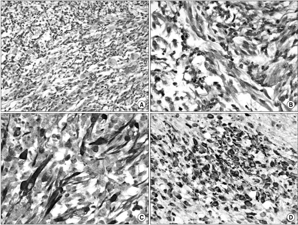
Fig. 3 Microscopic examination with H&E and immunohistochemical staining. (A) The tumor is composed predominantly of primitive ovoid cells with scattered rabdomyoblasts. The rhabdomyoblast in this case have eccentric vesicular nuclei and abundant densely eosinophilic cytoplasm (H&E, ×200), (B) elongated rhabdomyoblasts with distinct cross-striations in eosinophilic cytoplasm (H&E, ×400), (C) diffuse, strong positive immunoreactivity for desmin (desmin stain, ×400), (D) diffuse, strong positive immunoreactivity for MyoD1 (MyoD1 stain, ×400).
Reference
-
1. Kim DI, Kim KS. Two cases of pediatric paratesticular rhabdomyosarcoma. Korean J Urol. 2004. 45:1072–1076.2. Qualman S, Lynch J, Bridge J, Parham D, Teot L, Meyer W, et al. Prevalence and clinical impact of anaplasia in childhood rhabdomyosarcoma: a report from the Soft Tissue Sarcoma Committee of the Children's Oncology Group. Cancer. 2008. 113:3242–3247.3. Aquino MR, Gibson DP, Bloom DA. Paratesticular rhabdomyosarcoma with metastatic encasement of the abdominal aorta. Pediatr Radiol. 2011. 41:1061–1064.
Article4. Mak CW, Chou CK, Su CC, Huan SK, Chang JM. Ultrasound diagnosis of paratesticular rhabdomyosarcoma. Br J Radiol. 2004. 77:250–252.
Article5. Akbar SA, Sayyed TA, Jafri SZ, Hasteh F, Neill JS. Multimodality imaging of paratesticular neoplasms and their rare mimics. Radiographics. 2003. 23:1461–1476.
Article6. Raney RB, Anderson JR, Barr FG, Donaldson SS, Pappo AS, Qualman SJ, et al. Rhabdomyosarcoma and undifferentiated sarcoma in the first two decades of life: a selective review of intergroup rhabdomyosarcoma study group experience and rationale for Intergroup Rhabdomyosarcoma Study V. J Pediatr Hematol Oncol. 2001. 23:215–220.
Article7. Chung JM, Lim YT, Lee SD. Infantile testicular rhabdomyosarcoma. Urology. 2007. 69:1208.
Article
