Idiopathic Pleuroparenchymal Fibroelastosis Presenting in Recurrent Pneumothorax: A Case Report
- Affiliations
-
- 1Department of Internal Medicine, The Catholic University of Korea College of Medicine, Seoul, Korea. jssong@catholic.ac.kr
- 2Department of Pathology, The Catholic University of Korea College of Medicine, Seoul, Korea.
- 3Department of Radiology, The Catholic University of Korea College of Medicine, Seoul, Korea.
- KMID: 2320576
- DOI: http://doi.org/10.4046/trd.2014.77.4.184
Abstract
- Idiopathic pleuroparenchymal fibroelastosis (PPFE) is a rare, recently classified entity that consists of pleural and subjacent parenchymal fibrosis predominantly in the upper lungs. In an official American Thoracic Society/European Respiratory Society statement in 2013, this disease is introduced as a group of rare idiopathic interstitial pneumonias. We describe a case of a 76-year-old woman with cough and recurrent pneumothorax. She was admitted to our hospital with severe cough at first. High resolution computed tomography (HRCT) disclosed multifocal subpleural consolidations with reticular opacities in both lungs, primarily in the upper lobes, suggesting interstitial pneumonia. Rheumatoid lung was diagnosed initially through an elevated rheumatoid factor, HRCT and surgical biopsy at the right lower lobe. However, one month later, pneumothorax recurred. Surgical biopsy was performed at the right upper lobe at this time. The specimens revealed typical subpleural fibroelastosis. We report this as a first case of idiopathic PPFE in Korea after reviewing the symptoms, imaging and pathologic findings.
MeSH Terms
Figure
-

Figure 1 Chest radiograph shows multifocal consolidations and reticulonodular infiltrations in peripheral portion of both lungs. These lesions show upper lobe predominancy. Small pneumothorax is noted in right lung.
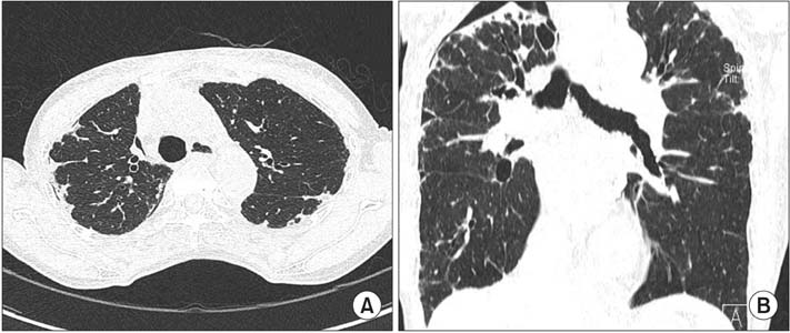
Figure 2 (A) High resolution computed tomography (HRCT) shows multifocal subpleural consolidations with reticular opacities in both lungs and small pneumothorax in right lung. Architectural distortions and traction bronchiectasis are combined in both lungs. (B) Coronal HRCT shows that these lesions predominantly affect the upper lobes.

Figure 3 Surgical lung biopsy specimen (H&E stain, ×40) at low power demonstrated dense fibrosis in the subpleura, lung parenchyme, and interstitium with temporal heterogeneity.
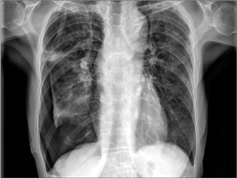
Figure 4 A chest radiagraph shows an increased amount of pneumothorax in the right lung compared with Figure 1.
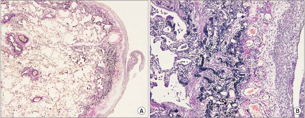
Figure 5 Surgical lung biopsy specimen (Elastic Van Gieson stain) at low power (A, ×100) shows pleural thickening and subpleural fibrosis. High power (B, ×200) demonstrated dense masses of elastic fibers beneath the thickend pleura.
Reference
-
1. Frankel SK, Cool CD, Lynch DA, Brown KK. Idiopathic pleuroparenchymal fibroelastosis: description of a novel clinicopathologic entity. Chest. 2004; 126:2007–2013.2. Becker CD, Gil J, Padilla ML. Idiopathic pleuroparenchymal fibroelastosis: an unrecognized or misdiagnosed entity? Mod Pathol. 2008; 21:784–787.3. Travis WD, Costabel U, Hansell DM, King TE Jr, Lynch DA, Nicholson AG, et al. An official American Thoracic Society/European Respiratory Society statement: Update of the international multidisciplinary classification of the idiopathic interstitial pneumonias. Am J Respir Crit Care Med. 2013; 188:733–748.4. Reddy TL, Tominaga M, Hansell DM, von der Thusen J, Rassl D, Parfrey H, et al. Pleuroparenchymal fibroelastosis: a spectrum of histopathological and imaging phenotypes. Eur Respir J. 2012; 40:377–385.5. Piciucchi S, Tomassetti S, Casoni G, Sverzellati N, Carloni A, Dubini A, et al. High resolution CT and histological findings in idiopathic pleuroparenchymal fibroelastosis: features and differential diagnosis. Respir Res. 2011; 12:111.6. von der Thusen JH, Hansell DM, Tominaga M, Veys PA, Ashworth MT, Owens CM, et al. Pleuroparenchymal fibroelastosis in patients with pulmonary disease secondary to bone marrow transplantation. Mod Pathol. 2011; 24:1633–1639.
- Full Text Links
-
- Actions
-
Cited
- CITED
-
- Close
- Share
-
- Similar articles
-
- Idiopathic Pleuroparenchymal Fibroelastosis, a Rare Entity of Interstitial Pneumonia: A Case Report
- Pleural Metastasis of Lung Cancer Combined with Pleuroparenchymal Fibroelastosis: A Case Report
- Pleuroparenchymal fibroelastosis in Korean patients: clinico-radiologic-pathologic features and 2-year follow-up
- Recurrent Pneumothorax Caused by an Unexpected Lymphangioleiomyomatosis: A Case Report
- Hemitruncus arteriosus associated with Endocardial Fibroelastosis
