A Case of Pulmonary Sarcoidosis with Endobronchial Nodular Involvement
- Affiliations
-
- 1Department of Internal Medicine, Wonkwang University College of Medicine, Iksan, Korea. yshpul@wku.ac.kr
- 2Department of Radiology, Wonkwang University College of Medicine, Iksan, Korea.
- 3Department of Pathology, Wonkwang University College of Medicine, Iksan, Korea.
Abstract
- Sarcoidosis is a multisystemic disorder of unknown cause that is characterized pathologically by noncaseating granulomas. Diagnosis is based on the exclusion of other infectious, interstitial, and neoplastic diseases and on the typical pathology. Although the lungs and mediastinal lymph nodes are almost involved, endobronchial nodular lesions of sarcoidosis with lung involvements are rare. We report a case of sarcoidosis with lung involvements and endobronchial nodules as confirmed by bronchial biopsy.
Keyword
Figure
-
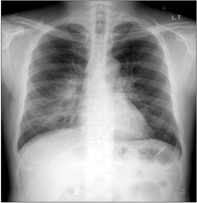
Figure 1 Chest radiograph showing ill-defined nodular peribronchial lesions in both lungs, most prominently in the mid right and lower lung zones.
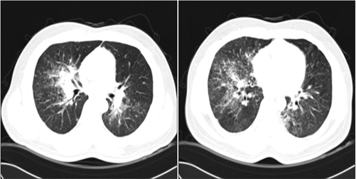
Figure 2 Chest computed tomography showed consolidation along the right upper and intermediate bronchus and small perilymphatic, centrilobular, and bronchovascular nodules with interlobular septal thickening in the upper right, mid, and lower lobes and the lower left lobe.

Figure 3 Follow-up chest radiography and computed tomography showed an increased extent of peribronchial infiltration and air space consolidation, interlobular septal thickening, and ill-defined nodules in both lungs, along with increased size of the multiple enlarged hilar and interlobar lymph nodes.
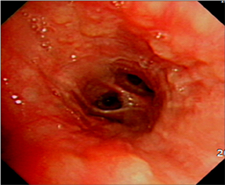
Figure 4 Bronchoscopy showed multiple variable small nodular endobronchial lesions.
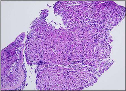
Figure 5 Biopsy of endobronchial nodules showed diffuse granulomatous inflammation with multinucleated giant cells (H&E stain, ×100).
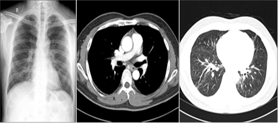
Figure 6 Follow-up chest radiography and computed tomography after treatment with oral steroid showed decreased extent of the peribronchial infiltration and air space consolidation in both lungs, along with a significantly decreased extent of the interlobar septal thickening and perilymphatic or ill-defined nodules, and decreased sizes of the multiple enlarged hilar and interlobar lymph nodes.
Reference
-
1. Iannuzzi MC, Rybicki BA, Teirstein AS. Sarcoidosis. N Engl J Med. 2007; 357:2153–2165.2. Kim DS, Ahn JJ. Sacoidosis in Korea. Tuberc Respir Dis. 2000; 49:274–280.3. Newman LS, Rose CS, Maier LA. Sarcoidosis. N Engl J Med. 1997; 336:1224–1234.4. Choi HH, Hong YA, Choi JK, Kim JS, Kim SJ, Kim SC, et al. A case of sarcoidosis that was initially misdiagnosed as nontuberculous mycobacteria pulmonary disease. Tuberc Respir Dis. 2009; 66:309–313.5. Hanno R, Callen JP. Sarcoidosis: a disorder with prominent cutaneous features and their interrelationship with systemic disease. Med Clin North Am. 1980; 64:847–866.6. Sharma OP. Sarcoidosis: a worldwide phenomenon. Sarcoidosis. 1984; 1:11–15.7. Nam JE, Ryu YH, Park JG, Choe KO, Im JG, Lee KS, et al. High resolution CT findings of pseudoalveolar sarcoidosis. J Korean Radiol Soc. 2002; 47:191–196.8. Tay HL, Vaughan-Jones R, Qureshi SS. Ethmoidal sarcoidosis. J Laryngol Otol. 1994; 108:682–684.9. Nishimura K, Itoh H, Kitaichi M, Nagai S, Izumi T. Pulmonary sarcoidosis: correlation of CT and histopathologic findings. Radiology. 1993; 189:105–109.10. Lee SJ, Kim JY, Lee JC, Kim GS, Yoo CG, Kim YW, et al. A case of sarcoidosis involving bone marrow, skin, uvea, joints, liver. Korean J Med. 1997; 53:580–585.11. Chung SG, Park CH. Axillary lymph node sarcoidosis. J Korean Surg Soc. 2001; 61:220–223.12. Lee BH, Kim JM, Kim DW, Kim JH, Bang KT, Lee KY, et al. A case of sarcoidosis with cavitation. Tuberc Respir Dis. 2005; 59:546–550.
