Quality of root canal fillings using three gutta-percha obturation techniques
- Affiliations
-
- 1Government Dental Service, Department of Health, the Government of Hong Kong SAR, Hong Kong, HKSAR, China.
- 2Area of Endodontics, Faculty of Dentistry, University of Hong Kong, Saiyingpun, Hong Kong, HKSAR, China. spcheung@hku.hk
- KMID: 2316960
- DOI: http://doi.org/10.5395/rde.2016.41.1.22
Abstract
OBJECTIVES
The goal of this study was to compare the density of gutta-percha root fillings obturated with the following techniques: cold lateral (CL) compaction, ultrasonic lateral (UL) compaction, and warm vertical (WV) compaction.
MATERIALS AND METHODS
Thirty-three extracted mandibular first molars, with two separate mesial canals in each, were selected. After instrumentation, the canals were stratified into three groups based on canal length and curvature, and underwent obturation with one of the techniques. No sealer was used in order to avoid masking any voids. The teeth were imaged pre- and post-obturation using micro-computed tomography. The reconstructed three-dimensional images were analyzed volumetrically to determine the amount of gutta-percha present in every 2 mm segment of the canal. P values < 0.05 were considered to indicate statistical significance.
RESULTS
The overall mean volume fraction of gutta-percha was 68.51 +/- 6.75% for CL, 86.56 +/- 5.00% for UL, and 88.91 +/- 5.16% for WV. Significant differences were found between CL and UL and between CL and WV (p < 0.05), but not between UL and WV (p = 0.526). The gutta-percha density of the roots treated with WV and UL increased towards the coronal aspect, but this trend was not noted in the CL group.
CONCLUSIONS
WV compaction and UL compaction produced a significantly denser gutta-percha root filling than CL compaction. The density of gutta-percha was observed to increase towards the coronal aspect when the former two techniques were used.
Keyword
MeSH Terms
Figure
-
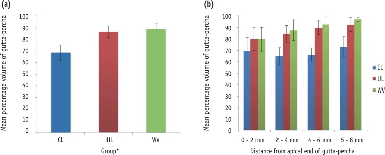
Figure 1 (a) Volume fraction of gutta-percha present in the root canal using cold lateral compaction, ultrasonic lateral compaction, and warm vertical compaction over the entire canal. The variation in this volume fraction value along the length of the root canal is shown in (b). CL, cold lateral compaction; UL, ultrasound lateral compaction; WV, warm vertical compaction.
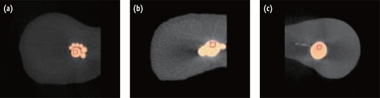
Figure 2 The micro-computed tomography appearance of selected root canals obturated using (a) cold lateral compaction, (b) ultrasonic lateral compaction, and (c) warm vertical compaction.
Cited by 3 articles
-
Micro-computed tomographic evaluation of a new system for root canal filling using calcium silicate-based root canal sealers
Mario Tanomaru-Filho, Fernanda Ferrari Esteves Torres, Jader Camilo Pinto, Airton Oliveira Santos-Junior, Karina Ines Medina Carita Tavares, Juliane Maria Guerreiro-Tanomaru
Restor Dent Endod. 2020;45(3):e34. doi: 10.5395/rde.2020.45.e34.Quantification of the tug-back by measuring the pulling force and micro computed tomographic evaluation
Su-Jin Jeon, Young-Mi Moon, Min-Seock Seo
Restor Dent Endod. 2017;42(4):273-281. doi: 10.5395/rde.2017.42.4.273.Influence of the root canal filling technique on the success rate of primary endodontic treatments: a systematic review
Daniel Feijolo Marconi, Giovana Siocheta da Silva, Theodoro Weissheimer, Isadora Ames Silva, Gabriel Barcelos Só, Leonardo Thomasi Jahnke, Jovito Adiel Skupien, Marcus Vinicius Reis Só, Ricardo Abreu da Rosa
Restor Dent Endod. 2022;47(4):e40. doi: 10.5395/rde.2022.47.e40.
Reference
-
1. Schilder H. Filling root canals in three dimensions. Dent Clin North Am. 1967; 11:723–744. PMID: 5262492.
Article2. Lea CS, Apicella MJ, Mines P, Yancich PP, Parker MH. Comparison of the obturation density of cold lateral compaction versus warm vertical compaction using the continuous wave of condensation technique. J Endod. 2005; 31:37–39. PMID: 15614003.
Article3. Gulabivala K, Holt R, Long B. An in vitro comparison of thermoplasticised gutta-percha obturation techniques with cold lateral condensation. Endod Dent Traumatol. 1998; 14:262–269. PMID: 9972158.4. Whitworth J. Methods of filling root canals: principles and practices. Endod Topics. 2005; 12:2–24.
Article5. Collins J, Walker MP, Kulild J, Lee C. A comparison of three gutta-percha obturation techniques to replicate canal irregularities. J Endod. 2006; 32:762–765. PMID: 16861078.
Article6. Goldberg F, Artaza LP, De Silvio A. Effectiveness of different obturation techniques in the filling of simulated lateral canals. J Endod. 2001; 27:362–364. PMID: 11485258.
Article7. Wong M, Peters DD, Lorton L. Comparison of gutta-percha filling techniques, compaction (mechanical), vertical (warm), and lateral condensation techniques, Part 1. J Endod. 1981; 7:551–558. PMID: 6985547.
Article8. Holcomb JQ, Pitts DL, Nicholls JI. Further investigation of spreader loads required to cause vertical root fracture during lateral condensation. J Endod. 1987; 13:277–284. PMID: 3474346.
Article9. Saw LH, Messer HH. Root strains associated with different obturation techniques. J Endod. 1995; 21:314–320. PMID: 7673840.
Article10. Moreno A. Thermomechanically softened gutta-percha root canal filling. J Endod. 1977; 3:186–188. PMID: 266028.
Article11. Baumgardner KR, Krell KV. Ultrasonic condensation of gutta-percha: an in vitro dye penetration and scanning electron microscopic study. J Endod. 1990; 16:253–259. PMID: 2074421.12. Deitch AK, Liewehr FR, West LA, Patton WR. A comparison of fill density obtained by supplementing cold lateral condensation with ultrasonic condensation. J Endod. 2002; 28:665–667. PMID: 12236312.
Article13. Bailey GC, Ng YL, Cunnington SA, Barber P, Gulabivala K, Setchell DJ. Root canal obturation by ultrasonic condensation of gutta-percha. Part II: an in vitro investigation of the quality of obturation. Int Endod J. 2004; 37:694–698. PMID: 15347294.14. Bailey GC, Cunnington SA, Ng YL, Gulabivala K, Setchell DJ. Ultrasonic condensation of gutta-percha: the effect of power setting and activation time on temperature rise at the root surface - an in vitro study. Int Endod J. 2004; 37:447–454. PMID: 15189433.15. Sweatman TL, Baumgartner JC, Sakaguchi RL. Radicular temperatures associated with thermoplasticized gutta-percha. J Endod. 2001; 27:512–515. PMID: 11501588.
Article16. Rhodes JS, Ford TR, Lynch JA, Liepins PJ, Curtis RV. Micro-computed tomography: a new tool for Quality of three obturation techniques experimental endodontology. Int Endod J. 1999; 32:165–170. PMID: 10530203.17. Fan B, Cheung GS, Fan M, Gutmann JL, Bian Z. C-shaped canal system in mandibular second molars: part I - anatomical features. J Endod. 2004; 30:899–903. PMID: 15564874.18. Peters OA, Schönenberger K, Laib A. Effects of four Ni-Ti preparation techniques on root canal geometry assessed by micro-computed tomography. Int Endod J. 2001; 34:221–230. PMID: 12193268.19. Zogheib C, Naaman A, Sigurdsson A, Medioni E, Bourbouze G, Arbab-Chirani R. Comparative microcomputed tomographic evaluation of two carrier-based obturation systems. Clin Oral Investig. 2013; 17:1879–1883.
Article20. Somma F, Cretella G, Carotenuto M, Pecci R, Bedini R, De Biasi M, Angerame D. Quality of thermoplasticized and single point root fillings assessed by microcomputed tomography. Int Endod J. 2011; 44:362–369. PMID: 21255040.
Article21. Gandolfi MG, Parrilli AP, Fini M, Prati C, Dummer PM. 3D micro-CT analysis of the interface voids associated with Thermafil root fillings used with AH Plus or a flowable MTA sealer. Int Endod J. 2013; 46:253–263. PMID: 23039158.
Article22. Zaslansky P, Fratzl P, Rack A, Wu MK, Wesselink PR, Shemesh H. Identification of root filling interfaces by microscopy and tomography methods. Int Endod J. 2011; 44:395–401. PMID: 21219359.
Article23. Pruett JP, Clement DJ, Carnes DL Jr. Cyclic fatigue testing of nickel-titanium endodontic instruments. J Endod. 1997; 23:77–85. PMID: 9220735.
Article24. Chandler NP. Root canal filling. In : Chong BS, editor. Harty's endodontics in clinical practice. 6th ed. Edinburgh: Elsevier;2010. p. 131–157.25. Ruddle CJ. Three-dimensional obturation of the root canal system. Dent Today. 1992; 11:28–33. PMID: 1535776.26. Wu MK, Van der Sluis LW, Wesselink PR. A preliminary study of the percentage of gutta-percha-filled area in the apical canal filled with vertically compacted warm gutta-percha. Int Endod J. 2002; 35:527–535. PMID: 12190910.
Article27. Smith RS, Weller RN, Loushine RJ, Kimbrough WF. Effect of varying the depth of heat application on the adaptability of gutta-percha during warm vertical compaction. J Endod. 2000; 26:668–672. PMID: 11469298.
Article28. Wu MK, Wesselink PR. A primary observation on the preparation and obturation of oval canals. Int Endod J. 2001; 34:137–141. PMID: 11307262.
Article29. Brayton SM, Davis SR, Goldman M. Gutta-percha root canal fillings. An in vitro analysis. I. Oral Surg Oral Med Oral Pathol. 1973; 35:226–231. PMID: 4574392.
- Full Text Links
-
- Actions
-
Cited
- CITED
-
- Close
- Share
-
- Similar articles
-
- Obturation efficiency of non-standardized gutta-percha cone in curved root canals prepared with 0.06 taper nickel-titanium instruments
- Comparison of warm gutta-percha condensation techniques in ribbon shaped canal: weight of filled gutta-percha
- The effect of different confluence confirmation strategies on the obturation of Vertucci type II canal: micro-CT analysis
- Comparison of apical seal with or without the use of dentin adhesive system
- Influence of plugger penetration depth on the area of the canal space occupied by gutta-percha
