Restor Dent Endod.
2012 May;37(2):114-118.
Pre-prosthetic minor tooth movement with elastic separating ring & provisional restoration modification: case report
- Affiliations
-
- 1Department of Conservative Dentistry, Yonsei University College of Dentistry, Seoul, Korea. chanyoungl@yuhs.ac
Abstract
- Proximal caries or coronal defect in posterior teeth may result in the loss of proximal space and drifting of neighboring teeth, which makes restoration difficult. Inability to restore proper contours and to align tooth axis properly are commonly encountered problems when planning tooth restoration. Moreover, tilted teeth aggravate periodontal tissue breakdown, such as pseudo-pocket, and angular osseous defect. The purpose of this case presentation is to describe a simple technique for inducing minor tooth movement with orthodontic separating ring and provisional restoration modification. This method was used to create crown placement space on mesially tilted molar. This method is easy, simple and efficient technique which could be used in interproximal space gaining in selected situation.
Figure
-
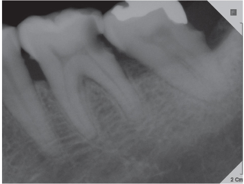
Figure 1 Preoperative periapical x-ray.
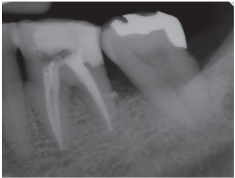
Figure 2 Periapical x-ray after root canal treatment.

Figure 3 Initial crown preparation prior to tooth movement.

Figure 4 Elastic ring inserted on #36, 37 interproximal area.
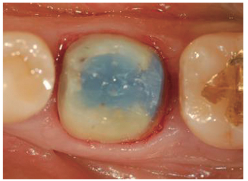
Figure 5 Final crown preparation after finishing tooth movement.
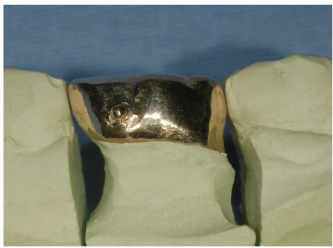
Figure 6 Die adaptation of cast gold crown. Note the recovery of proper distal proximal contour.
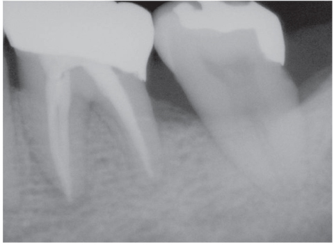
Figure 7 Postoperative periapical x-ray.

Figure 8 Postoperative clinical photo.
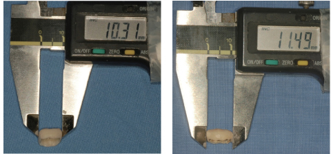
Figure 9 Estimate the amount of space gaining by measuring change in MD diameter of provisional crown. MD, mesio-distal.
Reference
-
1. Diedrich P. Preprosthetic orthodontics. J Orofac Orthop. 1996. 57:102–116.2. Mihram WL, Nemetz H. The tilted molar: a prosthetic and periodontal dilemma. Oral Health. 1991. 81:11–15.3. Keesee SM, Baty DL, Cameron SM, Lefler TB, Morris WJ. A technique for achieving prerestorative minor tooth movement with orthodontic separators. J Prosthet Dent. 2002. 88:544–547.
Article4. Smidt A, Venezia E. Gaining adequate interdental space with modified elastic separating rings: rationale and technique. Quintessence Int. 2002. 33:409–414.5. VanderWeele RA, Broome JC, Ramer JP. Regaining space by using elastic orthodontic separators. Gen Dent. 1998. 46:454–456.6. Davidovitch M, Papanicolaou S, Vardimon AD, Brosh T. Duration of elastomeric separation and effect on interproximal contact point characteristics. Am J Orthod Dentofacial Orthop. 2008. 133:414–422.
Article7. Bondemark L, Fredriksson K, Ilros S. Separation effect and perception of pain and discomfort from two types of orthodontic separators. World J Orthod. 2004. 5:172–176.8. Natali AN. Dental Biomechanics. 2003. London, UK and New York, USA: Taylor and Francis Group;196–197.9. Wehrbein H, Diedrich P. Mesio-marginal findings at tilted molars. A histological-histomorphometric study. Eur J Orthod. 2001. 23:663–670.
Article10. Kraal JH, Digiancinto JJ, Dail RA, Lemmerman K, Peden JW. Periodontal conditions in patients after molar uprighting. J Prosthet Dent. 1980. 43:156–162.
Article11. Lundgren D, Kurol J, Thorstensson B, Hugoson A. Periodontal conditions around tipped and upright molars in adults. An intra-individual retrospective study. Eur J Orthod. 1992. 14:449–455.
Article
- Full Text Links
-
- Actions
-
Cited
- CITED
-
- Close
- Share
-
- Similar articles
-
- Reinforcement of provisional restoration with cast metal framework: A case report
- Aesthetic prosthetic restoration through immediate implant placement and provisional restoration in the maxillary anterior region using a digital guide
- Decoronation and implant restoration of ankylosed tooth resulted from anterior avulsion: A case report
- Outcome Evaluation of an Immediately Placed Maxillary Anterior Single-Tooth Implant Using Objective Esthetic Criteria: Case Report
- Reinforcing the retention of provisional restoration using provisional implant on maxillary anterior region: clinical case report
