Korean J Urol.
2012 Nov;53(11):810-812.
Cavernous Hemangiolymphangioma of the Testis without Cutaneous Hemangiomatosis in an Elderly Patient
- Affiliations
-
- 1Department of Urology, Chonbuk National University Medical School, Jeonju, Korea. hjkim@jbnu.ac.kr
- 2Department of Anesthesiology, Chonbuk National University Medical School, Jeonju, Korea.
Abstract
- Hemangiolymphangioma is an extremely rare malformation of both the lymphatic and blood vessels. To date, however, there are no reports in the literature of a hemangiolymphangioma of the testis. An 84-year-old man visited our hospital for investigation of a 1-month episode of a rapidly growing mass in his right scrotum. Scrotal ultrasonography revealed a multilobulated mass with septation in the testis. Testicular tumor markers were within the normal limit. Radical orchiectomy was performed. At surgery, a red, wide-based, nodular tumor was found on the testis. Histological examination of the resected specimen showed it to be a cavernous hemangiolymphangioma. Here we report this first case of a cavernous hemangiolymphangioma of the testis without cutaneous hemangiomatosis in an elderly patient.
Keyword
MeSH Terms
Figure
-
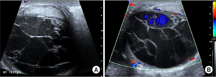
FIG. 1 (A) Scrotum ultrasonography showing a multilobulated mass with septation in the testis. (B) Low blood flow was seen on color Doppler ultrasonography.
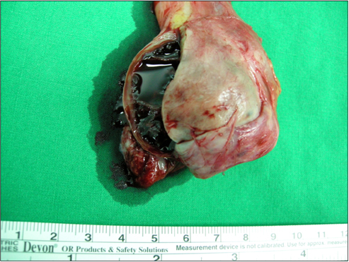
FIG. 2 Gross photograph of the testis after right radical orchiectomy. The testis was diffusely thickened and showed dilated blood vessels filled with blood.
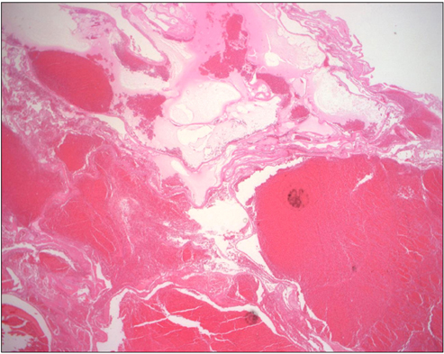
FIG. 3 Histopathological features of the hemangiolymphangioma showing both hemangiomatous and lymphangiomatous components (H&E, ×100).
Reference
-
1. Donnelly LF, Adams DM, Bisset GS 3rd. Vascular malformations and hemangiomas: a practical approach in a multidisciplinary clinic. AJR Am J Roentgenol. 2000. 174:597–608.2. Giacalone PL, Boulot P, Marty M, Deschamps F, Laffargue F, Viala JL. Fetal hemangiolymphangioma: a case report. Fetal Diagn Ther. 1993. 8:338–340.3. Paladini D, Lamberti A, Teodoro A, Liguori M, D'Armiento M, Capuano P, et al. Prenatal diagnosis and hemodynamic evaluation of Klippel-Trenaunay-Weber syndrome. Ultrasound Obstet Gynecol. 1998. 12:215–217.4. Tseng JJ, Chou MM, Ho ES. Fetal axillary hemangiolymphangioma with secondary intralesional bleeding: serial ultrasound findings. Ultrasound Obstet Gynecol. 2002. 19:403–406.5. Vilanova JC, Barcelo J, Smirniotopoulos JG, Perez-Andres R, Villalon M, Miro J, et al. Hemangioma from head to toe: MR imaging with pathologic correlation. Radiographics. 2004. 24:367–385.6. Cannard L, Lemelle JL, Gaconnet E, Champigneulle J, Mainard L, Claudon M. Dynamic MR imaging of bladder haemangioma. Pediatr Radiol. 2001. 31:882–885.7. Engel JD, Kuzel TM, Moceanu MC, Oefelein MG, Schaeffer AJ. Angiosarcoma of the bladder: a review. Urology. 1998. 52:778–784.8. Park JS, Chung DY, Kim SJ, Kim YS, Lee EJ, Park KH. Hemangioma of scrotum: a report of 3 cases. Korean J Urol. 1997. 38:885–888.9. Loberant N, Chernihovski A, Goldfeld M, Sweed Y, Vais M, Tzilman B, et al. Role of Doppler sonography in the diagnosis of cystic lymphangioma of the scrotum. J Clin Ultrasound. 2002. 30:384–387.10. Hamada Y, Yagi K, Tanano A, Kato Y, Takada K, Sato M, et al. Cystic lymphangioma of the scrotum. Pediatr Surg Int. 1998. 13:442–444.
- Full Text Links
-
- Actions
-
Cited
- CITED
-
- Close
- Share
-
- Similar articles
-
- Intraosseous hemangiolymphangioma of the mandible: a case report
- A Case of Acquired Multiple Cavernous Hemangiomatosis
- A Case of pulmonary cavernous hemangiomatosis presented with right shoulder pain
- Diffuse hepatic hemangiomatosis in an adult
- Benign Neonatal Hemagiomatosis with Unusually Persistent Lesions and Conjunctival Hemangioma
