Prospective Evaluation of the Accuracy of MDCT Angiography for Living Kidney Donor
- Affiliations
-
- 1Department of Urology, Urological Science Institute, Yonsei University Health System, Seoul, Korea. hanwk@yuhs.ac
- 2Department of Radiology, Yonsei University Health System, Seoul, Korea.
Abstract
- PURPOSE
In donor nephrectomy, it is important to understand the exact anatomy of the blood vessels during minimally invasive surgery. We prospectively analyzed the accuracy of the vessel structures obtained by use of 64-row multi-detector computed tomography (MDCT) angiography compared with the actual vessel structure observed during surgery.
MATERIALS AND METHODS
We analyzed 238 patients who underwent donor nephrectomy from July 2007 to August 2010. Before the operation, MDCT angiography was performed, and after the operation, the surgeons themselves wrote the protocol. The ipsilateral artery, the number of veins, the association with the run of the hilar vessel, and other vascular anomalies in computed tomography (CT) angiography and in the donor protocol were summarized.
RESULTS
Among 238 patients, nephrectomy was performed on the left side in 199 patients. The accuracy of MDCT for the artery and the vein was 93.3% and 92.4%, respectively. Accuracy did not differ significantly on the left and right sides (artery: p=0.124; vein: p=0.174). In 199 patients, the CT findings for the lumbar vein were compared with the surgical findings. The overall accuracy was shown to be 84.9%, and the accuracy of the group drained to the inferior vena cava (54%) was significantly different (p<0.01) from that of the group drained to the renal vein (98.6%). Thus, it may be necessary to pay close attention to the interpretation of the findings for the lumbar vein.
CONCLUSIONS
MDCT angiography is important for understanding the exact anatomy of blood vessels before minimally invasive surgery. We showed that 64-channel MDCT has high accuracy in the main vessel and hilar vessels. However, close attention to the interpretation of the CT findings for the lumbar vein may be required.
Keyword
MeSH Terms
Figure
-
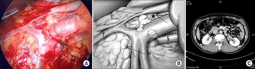
FIG. 1 (A) Actual image of the operation. (B) Structure with 1 renal artery (§) and 2 renal veins (★). 2 lumbar veins (†) merge and enter the lower renal vein. On the left side, the gonadal vein (*) is drained to the upper renal vein. The communication between the lumbar vein and the gonadal vein is shown. (C) Muti-detector computed tomography angiography of the same patient showing 1 renal artery and 2 renal veins. Lumbar veins were drained to the renal vein.
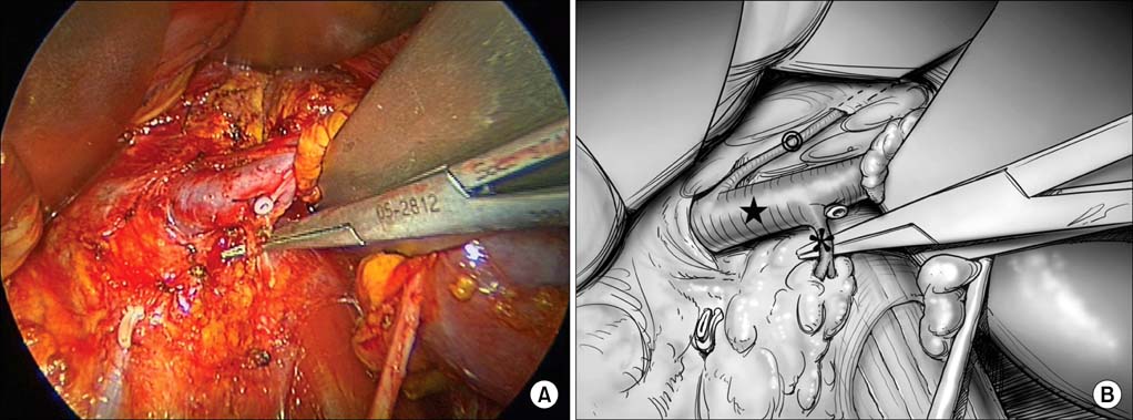
FIG. 2 (A) Actual image of the operation. (B) The lumbar vein (*) is drained to the renal vein (★), and the adrenal vein (◎) is drained to the renal vein.
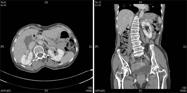
FIG. 3 Muti-detector computed tomography (MDCT) angiography image of a 60-year-old man. Although 1 renal artery and 1 renal vein (▾) were observed in the left kidney, an additional vein was found on the upper pole of the kidney during the actual operation.
Reference
-
1. Derauf B, Goldberg ME. Angiographic assessment of potential renal transplant donors. Radiol Clin North Am. 1987. 25:261–265.2. Shaffer D, Sahyoun AI, Madras PN, Monaco AP. Two hundred one consecutive living-donor nephrectomies. Arch Surg. 1998. 133:426–431.3. Shokeir AA, el-Diasty TA, Nabeeh A, Shaaban AA, el-Kenawy M, Wafa EW, et al. Digital subtraction angiography in potential live-kidney donors: a study of 1000 cases. Abdom Imaging. 1994. 19:461–465.4. Walker TG, Geller SC, Delmonico FL, Waltman AC, Athanasoulis CA. Donor renal angiography: its influence on the decision to use the right or left kidney. AJR Am J Roentgenol. 1988. 151:1149–1151.5. Sussman SK, Weinerth JL, Braun SD, Saeed M, Illescas FF, Cohan RH, et al. Intravenous digital subtraction angiography in the evaluation of potential renal donors. J Urol. 1987. 138:28–32.6. Kawamoto S, Montgomery R, Lawler LP, Horton KM, Fishman EK. Multidetector CT angiography for preoperative evaluation of living laparoscopic kidney donors. AJR Am J Roentgenol. 2003. 180:1633–1638.7. Rubin GD, Alfrey EJ, Dake MD, Semba CP, Sommer FG, Kuo PC, et al. Assessment of living renal donors with spiral CT. Radiology. 1995. 195:457–462.8. Pozniak MA, Balison DJ, Lee FT Jr, Tambeaux RH, Uehling DT, Moon TD. CT angiography of potential renal transplant donors. Radiographics. 1998. 18:565–587.9. el-Diasty TA, Shokeir AA, el-Ghar ME, Gad HM, Refaie AF, el-Din AB. Contrast enhanced spiral computerized tomography in live kidney donors: a single session for anatomical and functional assessment. J Urol. 2004. 171:31–34.10. Kawamoto S, Montgomery RA, Lawler LP, Horton KM, Fishman EK. Multi-detector row CT evaluation of living renal donors prior to laparoscopic nephrectomy. Radiographics. 2004. 24:453–466.11. Rastogi N, Sahani DV, Blake MA, Ko DC, Mueller PR. Evaluation of living renal donors: accuracy of three-dimensional 16-section CT. Radiology. 2006. 240:136–144.12. Roh JR, Park CM, Hyun JH, Ryu JA, Kim B, Lee SI, et al. Preoperative evaluation of living renal transplant donors using helical CT angiography: comparison with conventional angiography. Korean J Urol. 2002. 43:43–48.13. Yang SC, Rha KH, Byun YJ, Kim WY. Video-assisted minilaparotomy in urology. J Endourol. 2003. 17:465–467.14. Schlunt LB, Harper JD, Broome DR, Baron PW, Watkins GE, Ojogho ON, et al. Improved detection of renal vascular anatomy using multidetector CT angiography: Is 100% detection possible? J Endourol. 2007. 21:12–17.15. Raman S, Pojchamarnwiputh S, Muangsomboon K, Schulam PG, Gritsch HA, Lu DS. Utility of 16-MDCT angiography for comprehensive preoperative vascular evaluation of laparoscopic renal donors. AJR Am J Roentgenol. 2006. 186:1630–1638.16. Ratner LE, Kavoussi LR, Chavin KD, Montgomery R. Laparoscopic live donor nephrectomy: technical considerations and allograft vascular length. Transplantation. 1998. 65:1657–1658.17. Lewis GR, Mulcahy K, Brook NR, Veitch PS, Nicholson ML. A prospective study of the predictive power of spiral computed tomographic angiography for defining renal vascular anatomy before live-donor nephrectomy. BJU Int. 2004. 94:1077–1081.18. Bhatti AA, Chugtai A, Haslam P, Talbot D, Rix DA, Soomro NA. Prospective study comparing three-dimensional computed tomography and magnetic resonance imaging for evaluating the renal vascular anatomy in potential living renal donors. BJU Int. 2005. 96:1105–1108.19. Choi KH, Lee JW, Rha KH, Yang SC, Han WK. Standardized retroperitoneal minilaparotomy live donor nephrectomy in consecutive 615 cases. Korean J Endourol. 2010. 9:Suppl 1. 132. abstract 5-1.20. Janschek EC, Rothe AU, Hölzenbein TJ, Langer F, Brugger PC, Pokorny H, et al. Anatomic basis of right renal vein extension for cadaveric kidney transplantation. Urology. 2004. 63:660–664.
- Full Text Links
-
- Actions
-
Cited
- CITED
-
- Close
- Share
-
- Similar articles
-
- CT Angiography for Living Kidney Donors: Accuracy, Cause of Misinterpretation and Prevalence of Variation
- Evaluation of the Recipient and Donor in Living Kidney Transplantation
- Gadolinium-enhanced MR Angiography in Living Donor Renal Transplantation
- Living Donor Nephrectomy
- MDCT Application for Coronary Artery Intervention
