CT Angiography for Living Kidney Donors: Accuracy, Cause of Misinterpretation and Prevalence of Variation
- Affiliations
-
- 1Department of Radiology, Seoul National University College of Medicine, The Institute of Radiation Medicine, Clinical Research Institute, Seoul National University Hospital, Seoul, Korea. leew@radiol.snu.ac.kr
- 2Department of Urology, Seoul National University College of Medicine, Seoul, Korea.
- KMID: 1076459
- DOI: http://doi.org/10.3348/kjr.2008.9.4.333
Abstract
OBJECTIVE
To determine the accuracy of the use of multi-detector row CT (MDCT) to predict vascular anatomy in living kidney donors and to reveal the prevalence of vascular variations in a Korean population. MATERIALS AND METHODS: A total of 153 living kidney donors that had undergone preoperative CT and nephrectomy, either with open or laparoscopic surgery, were selected retrospectively. The initial CT results were compared with the surgical findings and repeated review sessions of CT scans were performed to determine the causes of mismatches in discordant cases. RESULTS: The accuracy of CT angiography was 95% to predict the number of renal vessels. Four arteries and two veins were missed during the initial CT interpretation due to perception errors (for two arteries and two veins) and technical limitations (two arteries). The prevalence of multiple renal arteries and veins, early branching of a renal artery and late confluence of a renal vein were 31%, 5%, 12%, 17%, respectively. The circumaortic renal vein and the bilateral inferior vena cava were found in two cases each (1.3%). One case (0.7%) each of a retroaortic renal vein and a supradiaphragmatic originated renal artery were found. CONCLUSION: MDCT provides a reliable method to evaluate the vascular anatomy and variations of living kidney donors.
MeSH Terms
Figure
-
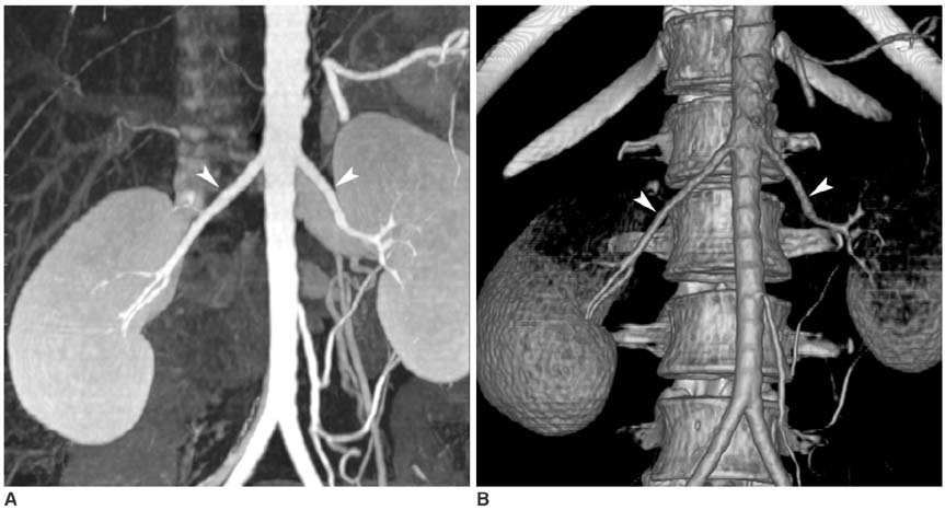
Fig. 1 31-year-old female, left kidney donor. A, B. Maximum intensity projection image and 3D volume rendered image show bilateral single renal arteries (arrowheads). Two left renal arteries were found during donor nephrectomy. However, retrospective review with knowledge of surgical results revealed only one visible renal artery.
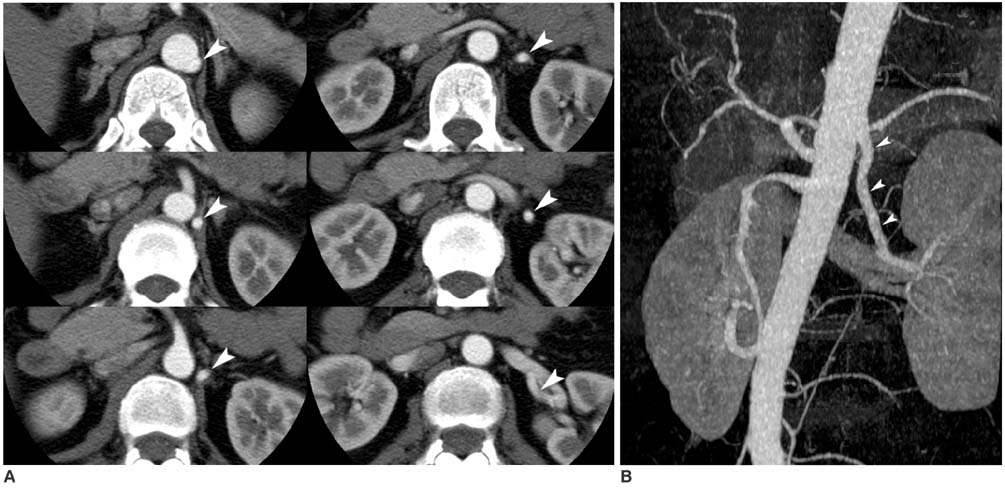
Fig. 2 49-year-old female, left kidney donor. A, B. Contrast-enhanced arterial phase axial CT scan images and maximum intensity projection image showing left renal artery with supradiaphragmatic origin (arrowheads in A, B), which is known as rare variation. In this case, left renal artery length was sufficient for donor nephrectomy.
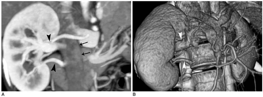
Fig. 3 23-year-old female, right kidney donor. One right renal vein was detected at initial CT interpretation, but two right renal veins were found during donor nephrectomy. A, B. Maximum intensity projection image and 3D volume rendered image show two right renal veins. Retrospective review without knowledge of surgical results revealed accessory renal vein (arrowheads in A, B) confluence at lower level of inferior vena cava (arrows in A).
Reference
-
1. Letourneau JG, Day DL, Ascher NL, Castaneda-Zuniga WR. Imaging of renal transplants. AJR Am J Roentgenol. 1988. 150:833–838.2. Smith PA, Ratner LE, Lynch FC, Corl FM, Fishman EK. Role of CT angiography in the preoperative evaluation for laparoscopic nephrectomy. Radiographics. 1998. 18:589–601.3. Dachman AH, Newmark GM, Mitchell MT, Woodle ES. Helical CT examination of potential kidney donors. AJR Am J Roentgenol. 1998. 171:193–200.4. Patil UD, Ragavan A, Nadaraj , Murthy K, Shankar R, Bastani B, et al. Helical CT angiography in evaluation of live kidney donors. Nephrol Dial Transplant. 2001. 16:1900–1904.5. Kawamoto S, Montgomery RA, Lawler LP, Horton KM, Fisherman EK. Multi-detector row CT evaluation of living renal donors prior to laparoscopic nephrectomy. Radiographics. 2004. 24:453–466.6. Holden A, Smith A, Dukes P, Pilmore H, Yasutomi M. Assessment of 100 live potential renal donors for laparoscopic nephrectomy with Multi-detector row helical CT. Radiology. 2005. 237:973–980.7. Kim JK, Park SY, Kim HJ, Kim CS, Ahn HJ, Ahn TY, et al. Living donor kidneys : usefulness of multi-detector row CT for comprehensive evaluation. Radiology. 2003. 229:869–876.8. Sahani DV, Rastogi N, Greenfield AC, Kalva SP, Ko D, Saini S, et al. Multi-detector row CT in evaluation of 94 living renal donors by readers with varied experience. Radiology. 2005. 235:905–910.9. Raman SS, Pojchamarnwiputh S, Muanqsomboon K, Schulam PG, Gritsch HA, Lu DS. Utility of 16-MDCT angiography for comprehensive preoperative vascular evaluation of laparoscopic renal donors. AJR Am J Roentgenol. 2006. 186:1630–1638.10. Namasivayam S, Small WC, Kalra MK, Torres WE, Newell KA, Mittal PK. Multidetector-row CT angiography for preoperative evaluation of potential laparoscopic renal donors: how accurate are we? Clin Imaging. 2006. 30:120–126.11. Villablanca JP, Rodriguez FJ, Stockman T, Dahliwal S, Omura M, Hazany S, et al. MDCT angiography for detection and quantification of small intracranial arteries:comparison with conventional catheter angiography. AJR Am J Roentgenol. 2007. 188:593–602.12. Satyapal KS, Haffejee AA, Singh B, Ramsaroop L, Robbs JV, Kalideen JM. Additional renal arteries : incidence and morphometry. Surg Radiol Anat. 2001. 23:33–38.13. Claves JL, Wise SW, Hopper KD, Tully D, Ten Have TR, Weaver J. Evaluation of contrast densities in the diagnosis of the carotid stenosis by CT angiography. AJR Am J Roentgenol. 1997. 169:569–573.14. Pozniak MA, Balison DJ, Lee FT Jr, Tambeaux RH, Uehling DT, Moon TD. CT angiography of potential renal transplant donors. Radiographics. 1998. 18:565–587.15. Rydberg J, Kopecky KK, Tann M, Persohn SA, Leapman SB, Filo RS, et al. Evaluation of prospective living renal donors for laparoscopic nephrectomy with multisection CT: the marriage of minimally invasive imaging with minimally invasive surgery. Radiographics. 2001. 21(Spec Issue):S223–S236.16. Neymark E, LaBerge JM, Hirose R, Melzer JS, Kerlan RK Jr, Wilson MW, et al. Arteriographic detection of renovascular disease in potential renal donors: incidence and effect on donor surgery. Radiology. 2000. 214:755–760.17. Urban BA, Ratner LE, Fishman EK. Three-dimensional volume-rendered CT angiography of the renal arteries and veins : normal anatomy, variants, and clinical applications. Radiographics. 2001. 21:373–386.
- Full Text Links
-
- Actions
-
Cited
- CITED
-
- Close
- Share
-
- Similar articles
-
- Prospective Evaluation of the Accuracy of MDCT Angiography for Living Kidney Donor
- Gadolinium-enhanced MR Angiography in Living Donor Renal Transplantation
- Preoperative Reno-vascular Evaluation of Living Renal Donors with Contrast Enhanced Magnetic Resonance Angiography
- Living Donor Nephrectomy
- Psychosocial Pre-Transplant Assessment of Living Kidney Donors
