Malignant Mesothelioma of the Spermatic Cord
- Affiliations
-
- 1Department of Urology, Catholic University of Daegu, School of Medicine, Daegu, Korea. jspark@cu.ac.kr
- 2Department of Pathology, Catholic University of Daegu, School of Medicine, Daegu, Korea.
Abstract
- Primary tumors arising from the spermatic cord are very rare. Mesothelioma derives from the mesothelial cells lining the serous membrane, such as the pleura, peritoneum, and tunica vaginalis of testis. Paratesticular malignant mesothelioma (MM), which usually presents as a hydrocele or intrascrotal mass, accounts for 0.3% to 1.4% of MMs. MMs of the spermatic cord account for less than 10% of paratesticular MMs. We report a case of MM of the spermatic cord in a 65-year-old man who primarily presented to the hospital with a left inguinal mass. Following the diagnosis after surgery, he was found to have a contralateral right inguinal mass and died in 6 months. Despite their rare occurrence in the spermatic cord, MMs need to be suspected, especially in patients with a history of asbestos exposure.
Keyword
MeSH Terms
Figure
-
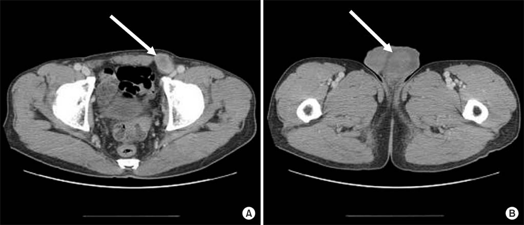
FIG. 1 Computed tomography scan showing a 3.2 cm lobulated mass in the left inguinal area (A) and a hydrocele in the left scrotum (B).
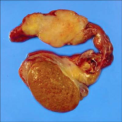
FIG. 2 On gross examination, the left orchiectomy specimen was composed of a 5.0×6.0 cm sized testis, attached to a 12 cm length spermatic cord and detached tunica vaginalis hydrocele sac. At the spermatic cord, there was a nodular mass measuring 5.0×3.2×2.5 cm. The outer surface of the mass was relatively well capsulated and the cut surface was shiny and whitish.
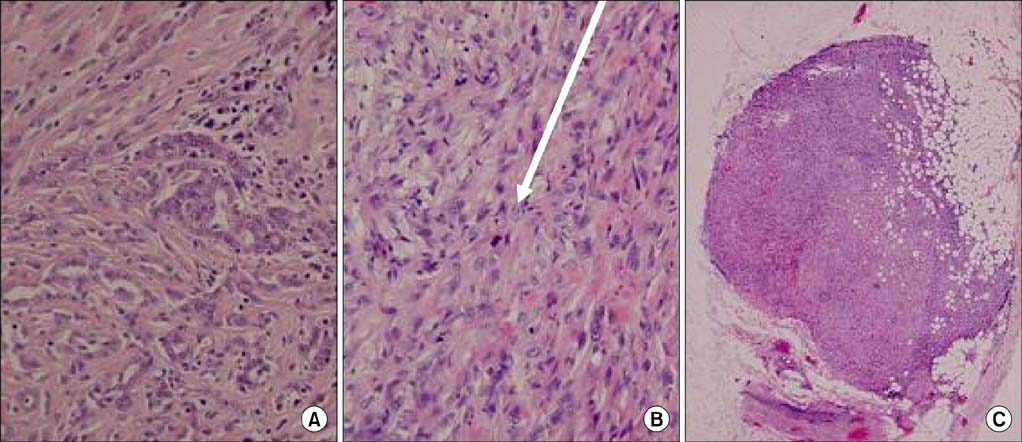
FIG. 3 (A) Epithelial tumor cells form a tubuloglandular structure (H&E, ×200). (B) Sarcomatous tumor cells composed of pleomorphic spindle cells with frequent abnormal mitosis (arrow) (H&E, ×200). (C) Peritoneal nodules show infiltrative sarcomatous tumor cells into the adipose tissue (H&E, ×40).
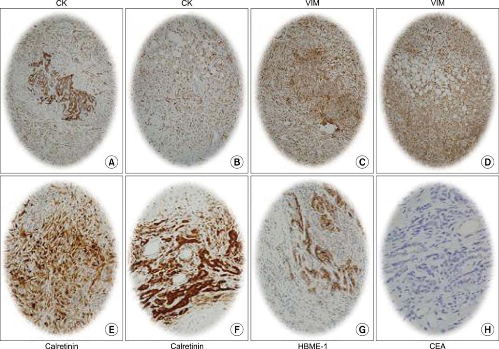
FIG. 4 The epithelial and sarcomatoid tumor cells were immunoreactive to cytokeratin (CK), calretinin, vimentin (VIM), and mesothelial antibody (HBME-1) but not to carcinoembryonic antigen (CEA) (immunochemical stain, ×100).
Reference
-
1. Park HM, Kim JH, Bae SG, Kwon TK, Chung SK. A case of malignant mesothelioma of tunica vaginalis. Korean J Urol. 1997. 38:1132–1134.2. Lee SC, Lee JK. A case of primary malignant mesothelioma of tunica vaginalis testis. Korean J Urol. 1991. 32:843–845.3. Shin TK, Lee TY, Park MH. Mesothelioma of the tunica vaginalis of the spermatic cord with coincidental finding renal cell carcinoma. Korean J Urol. 1995. 36:1142–1146.4. Plas E, Riedl CR, Pflüger H. Malignant mesothelioma of the tunica vaginalis testis: review of the literature and assessment of prognostic parameters. Cancer. 1998. 83:2437–2446.5. Perez-Ordonez B, Srigley JR. Mesothelial lesions of the paratesticular region. Semin Diagn Pathol. 2000. 17:294–306.6. Bisceglia M, Dor DB, Carosi I, Vairo M, Pasquinelli G. Paratesticular mesothelioma. Report of a case with comprehensive review of literature. Adv Anat Pathol. 2010. 17:53–70.7. Moore AJ, Parker RJ, Wiggins J. Malignant mesothelioma. Orphanet J Rare Dis. 2008. 3:34.8. García de Jalón A, Gil P, Azúa-Romeo J, Borque A, Sancho C, Rioja LA. Malignant mesothelioma of the tunica vaginalis. Report of a case without risk factors and review of the literature. Int Urol Nephrol. 2003. 35:59–62.9. Goel A, Agrawal A, Gupta R, Hari S, Dey AB. Malignant mesothelioma of the tunica vaginalis of the testis without exposure to asbestos. Cases J. 2008. 1:310.10. Hassan R, Alexander R. Nonpleural mesotheliomas: mesothelioma of the peritoneum, tunica vaginalis, and pericardium. Hematol Oncol Clin North Am. 2005. 19:1067–1087.
- Full Text Links
-
- Actions
-
Cited
- CITED
-
- Close
- Share
-
- Similar articles
-
- Mesothelioma of the Tunica Vaginalis of the Spermatic Cord with Coincidental Finding of Renal Cell Carcinoma
- Liposarcoma of Spermatic Cord: Report of A Case
- A Case of Rhabdomyosarcoma of Spermatic Cord
- A case of leiomyosarcoma of spermatic cord
- Clinical Consideration of Metastatic Tumors of the Spermatic Cord
