Korean J Urol.
2010 Jan;51(1):60-63.
The Clinical Significance of Intrarenal Reflux in Voiding Cystourethrography (VCUG)
- Affiliations
-
- 1Department of Urology, Urological Science Institute, Yonsei University College of Medicine, Seoul, Korea. swhan@yuhs.ac
Abstract
- PURPOSE
Intrarenal reflux (IRR) occurs in 3-10% of cases of total reflux and can lead to renal injury, which may eventually result in renal scarring. In this study, we evaluated the clinical importance of IRR detected by voiding cystourethrography and evaluated the relationship between IRR and renal scarring. MATERIALS AND METHODS: From January 2002 to May 2008, 50 patients who were diagnosed with vesicoureteral reflux (VUR) and showed IRR in voiding cystourethrography were enrolled. IRR was seen in 59 renal units in our enrolled patients. A 99mTc 2,3-dimercaptosuccinic acid (DMSA) renal scan was performed after VUR was detected in all cases. Nine patients were conservatively treated with prophylactic antibiotics, whereas 41 patients received an anti-reflux operation. A follow-up renal scan was performed after 3 to 6 months to check for any changes in renal scarring. RESULTS: The average patient age was 9.2 months (range, 1-42 months). Forty-nine patients were male; only one patient was female. The mean duration until surgery was 2.9 months. Generally, the IRR sites corresponded to the sites of photon defects on DMSA renal scans (76.3%). Furthermore, the photon defects on IRR sites tended to progress to renal scarring (65.2%). The rate of change in scarring was lower in the surgery group (47.1%) than in the prophylactic antibiotic group (75%). CONCLUSIONS: IRR sites and the sites of photon defects on DMSA renal scans showed a high correspondence, and these sites tended to progress to renal scarring. We suggest that VUR with IRR should be actively managed to decrease the chances of renal scarring.
MeSH Terms
Figure
-
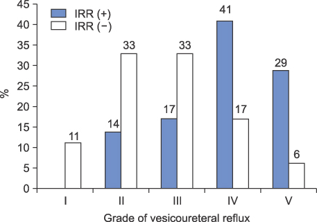
FIG. 1 Distributions of vesicoureteral reflux (VUR) grades according to the presence of intrarenal reflux (IRR).
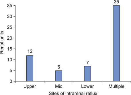
FIG. 2 Distribution of sites of intrarenal reflux (IRR) on voiding cystourethrography.
Reference
-
1. Gotoh T, Asano Y, Nonomura K, Togashi M, Koyanagi T. Intrarenal reflux in children with vesicoureteral reflux. Nippon Hinyokika Gakkai Zasshi. 1991. 82:1480–1486.2. Cremin BJ. Observations on vesico-ureteric reflux and intrarenal reflux: a review and survey of material. Clin Radiol. 1979. 30:607–621.3. Brodeur AE, Goyer RA, Melick W. A potential hazard of barium cystography. Radiology. 1965. 85:1080–1084.4. Bailey RR. The relationship of vesico-ureteric reflux to urinary tract infection and chronic pyelonephritis-reflux nephropathy. Clin Nephrol. 1973. 1:132–141.5. Darge K, Trusen A, Gordjani N, Riedmiller H. Intrarenal reflux: diagnosis with contrast-enhanced harmonic US. Pediatr Radiol. 2003. 33:729–731.6. Hodson CJ, Twohill SA. The time factor in the development of sterile renal scarring following high-pressure vesicoureteral reflux. Contrib Nephrol. 1984. 39:358–369.7. Hannerz L, Wikstad I, Johansson L, Broberger O, Aperia A. Distribution of renal scars and intrarenal reflux in children with a past history of urinary tract infection. Acta Radiol. 1987. 28:443–446.8. Ransley PG, Risdon RA. Reflux nephropathy: effects of antimicrobial therapy on the evolution of the early pyelonephritic scar. Kidney Int. 1981. 20:733–742.9. Ransley PG, Risdon RA, Godley ML. High pressure sterile vesicoureteral reflux and renal scarring: an experimental study in the pig and minipig. Contrib Nephrol. 1984. 39:320–343.10. Schulman SL, Snyder HM 3rd. Vesicoureteral reflux and reflux nephropathy in children. Curr Opin Pediatr. 1993. 5:191–197.11. Rolleston GL, Shannon FT, Utley WL. Relationship of infantile vesicoureteric reflux to renal damage. Br Med J. 1970. 1:460–463.12. Rolleston GL, Maling TM, Hodson CJ. Intrarenal reflux and the scarred kidney. Arch Dis Child. 1974. 49:531–539.13. Hodson CJ, Maling TM, McManamon PJ, Lewis MG. The pathogenesis of reflux nephropathy (chronic atrophic pyelonephritis). Br J Radiol. 1975. 13:Suppl 13. 1–26.14. Funston MR, Cremin BJ. Intrarenal reflux-papillary morphology and pressure relationships in children's necropsy kidneys. Br J Radiol. 1978. 51:665–670.15. Lim BT, Lee HS, Pai KS. Clinical significance of intrarenal reflux in children with urinary tract infection. J Korean Soc Pediatr Nephrol. 2008. 12:186–193.
- Full Text Links
-
- Actions
-
Cited
- CITED
-
- Close
- Share
-
- Similar articles
-
- Contrast-enhanced voiding urosonography for the diagnosis of vesicoureteral reflux and intrarenal reflux: a comparison of diagnostic performance with fluoroscopic voiding cystourethrography
- Clinical Significance of Mild Fetal Pelviectasia and The Role of Postnatal Voiding Cystourethrography
- The ABCs of Voiding Cystourethrography
- Value of the Voiding Cystourethrography Prior to Renal Transplantation
- Magnetic Resonance Voiding Cystography in the Diagnosis of Vesicoureteral Reflux: Comparative Study with Voiding Cystourethrography
