Korean J Urol.
2009 Mar;50(3):296-299.
Paraganglioma of a Bladder with a Bladder Stone
- Affiliations
-
- 1Department of Urology, College of Medicine, Dongguk University, Gyeongju, Korea. ksleemd@dongguk.ac.kr
- 2Department of Pathology, College of Medicine, Dongguk University, Gyeongju, Korea.
- 3Department of Internal Medicine, College of Medicine, Dongguk University, Gyeongju, Korea.
- 4Department of Radiology, College of Medicine, Dongguk University, Gyeongju, Korea.
Abstract
- Paragangliomas of the urinary bladder are very rare tumors of the paraganglion system that arise from chromaffin cells in or near the sympathetic ganglia. Only approximately 15% of them develop from extra-adrenal chromaffin tissue. Most of these tumors are hormonally active and secrete mainly noradrenaline (rarely adrenaline), calcitonin, and adrenocorticotropic hormone. Paragangliomas are generally benign tumors, with less than 10% being malignant. Here we report a case of a paraganglioma arising in a urinary bladder with a bladder stone.
MeSH Terms
Figure
-

Fig. 1 Pelvic CT scan showing a 2×2 cm sized calcification in the posterior wall of the bladder.

Fig. 2 Microscopic features of the tumor. The tumor was composed of nests of cuboidal epithelial cells surrounding a delicate vasculature. Tumor cells are characterized by relatively uniform round nuclei with fine chromatin and abundant amphophilic cytoplasm (H&E, ×200).
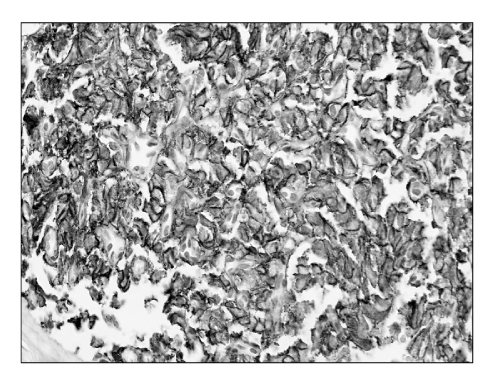
Fig. 3 Immunohistochemical stain for CD56. Tumor cells reveal a diffuse strong positive reaction to CD56 (×400).
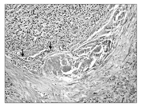
Fig. 4 Tumor infiltrates into the proper muscular layer (arrow)(H&E, ×100).
Reference
-
1. Siatelis A, Konstantinidis C, Volanis D, Leontara V, Thoma-Tsagli E, Delakas D. Pheochromocytoma of the urinary bladder: report of 2 cases and review of literature. Minerva Urol Nefrol. 2008. 60:137–140.2. Zimmerman IJ, Biron RE, MacMahon HE. Pheochromocytoma of the urinary bladder. N Engl J Med. 1953. 249:25–26.3. Seon IC, Kang SH, Kwak KM, Chung WK, Lee YS, Han CH, et al. Paraganglioma of the bladder. Korean J Urol. 2003. 44:198–200.4. Whalen RK, Althausen AF, Daniels GH. Extra-adrenal pheochromocytoma. J Urol. 1992. 147:1–10.5. Asbury WL Jr, Hatcher PA, Gould HR, Reeves WA, Wilson DD. Bladder pheochromocytoma with ring calcification. Abdom Imaging. 1996. 21:275–277.6. Onishi T, Sakata Y, Yonemura S, Sugimura Y. Pheochromocytoma of the urinary bladder without typical symptoms. Int J Urol. 2003. 10:398–400.7. Demirkesen O, Cetinel B, Yaycioglu O, Uygun N, Solok V. Usual cause of early preeclampsia: bladder paraganglioma. Urology. 2000. 56:154.8. Liu Y, Dong SG, Dong Z, Mao X, Shi XY. Diagnostic and treatment of pheochromocytoma in urinary bladder. J Zhejiang Univ Sci B. 2007. 8:435–438.9. Grignon DJ, Ro JY, Mackay B, Ordonez NG, el-Naggar A, Molina TJ, et al. Paraganglioma of the urinary bladder: immunohistochemical, ultrastructural, and DNA flow cytometric studies. Hum Pathol. 1991. 22:1162–1169.10. Singh DV, Seth A, Gupta NP, Kumar M. Calcified nonfunctional paraganglioma of the urinary bladder mistaken as bladder calculus: a diagnostic pitfall. BJU Int. 2000. 85:1152–1153.
- Full Text Links
-
- Actions
-
Cited
- CITED
-
- Close
- Share
-
- Similar articles
-
- Ultrasound, CT, and MR imaging Findings of Paraganglioma Originating at the Urinary Bladder: A Case Report
- Bladder performation due to foreigh body with stone in bladder: a case report
- Unusual Bladder Stones -Report of Three Cases-
- Foreign Body Induced Bladder Stone After Bladder Neck Suspension: A Case Report
- A Case of Intravesical Migration of Intrauterine Device with Stone Formation
