Korean J Urol.
2006 Feb;47(2):211-213. 10.4111/kju.2006.47.2.211.
Non-palpable and Asymptomatic Leydig Cell Tumor
- Affiliations
-
- 1Department of Urology, Soonchunhyang University College of Medicine, Seoul, Korea. yssong@hosp.sch.ac.kr
- 2Department of Pathology, Soonchunhyang University College of Medicine, Seoul, Korea.
- KMID: 2294204
- DOI: http://doi.org/10.4111/kju.2006.47.2.211
Abstract
- Leydig cell tumors are the most common non-germ cell tumors of the testis, and they account for 1-3% of all testicular tumors. They most commonly present as a testicular mass and/or with endocrine symptoms. This tumor is characterized by its endocrine manifestations, which are due to the tumor's capacity to secrete hormones. We report here on one case of a patient with Leydig cell tumor; the patient was without symptoms and the tumor was not detected on the physical examination.
Keyword
Figure
-
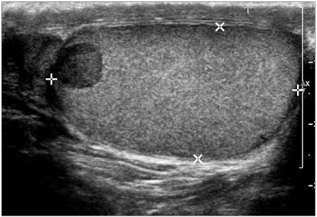
Fig. 1 The ultrasonographic findings show a 0.8×0.8cm sized hypoechogenic well marginated lesion in the upper portion of the right testis, which is confined within the tunica albuginea.
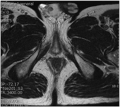
Fig. 2 T2 weighted axial image of the pelvic magnetic resonance imaging (MRI) shows a 1×1cm sized mass with low signal intensity, which is well marginated and has no infiltration beyond the tunica albuginea.

Fig. 3 Microscopic finding shows that the tumor is large and made of polygonal cells with abundant granular eosinophilic cytoplasm and prominent nucleoli (H&E, ×400).
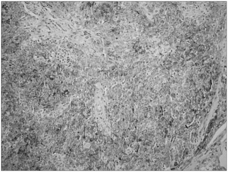
Fig. 4 Immunohistochemistry shows positive staining for α-inhibin (Inhibin, ×200).
Reference
-
1. Ritchey M. Walsh PC, Retik AB, Stamey TA, Vaughan ED, Wein AJ, editors. Pediatric urologic oncology. Campbell's urology. 2001. 8th ed. Philadelphia: Saunders;2498.2. Kim I, Yound RH, Scully RE. Leydig cell tumors of the testis. A clinicopathological analysis of 40 cases and review of the literature. Am J Surg Pathol. 1985. 9:177–192.3. Lee JJ, Park CG, Kim SJ, Shin KY, Park YW, Park HY, et al. A case of malignant Leydig cell tumor. Korean J Urol. 1997. 38:97–101.4. Na SS, Choi NG. A case of Leydig cell tumor of testis in child. Korean J Urol. 1988. 29:867–870.5. Cheon J, Shin YS, Cho JH, Kim SK, Kim IS. A case of malignant Leydig cell tumor of testis in adult. Korean J Urol. 1986. 27:349–355.6. Shim HY, Shin JH, Lee HY, Woo YN, Kim DH. A case of leydig cell tumor of testis in a 4-year-old boy. Korean J Urol. 1984. 25:242–246.7. Gabrilove JL, Nicolis GL, Mitty HA, Sohval AR. Feminizing interstitial cell tumor of the testis: personal observations and a review of the literature. Cancer. 1975. 35:1184–1202.8. Johnson DE, Swanson DA, von Eschenbach AC. Smith DR, editor. Tumors of the genitourinary tract. General urology. 1984. 11th ed. Los Altos, California: Lange;365.9. Caldamone AA, Altebarmakian V, Frank IN, Linke CA. Leydig cell tumor of testis. Urology. 1979. 14:39–43.10. van der Hem KG, Boven E, van Hennik MB, Pinedo HM. Malignant Leydig cell tumor of the testis in complete remission on o,p'-dichlorodiphenyl-dichloroethane. J Urol. 1992. 148:1256–1259.
