Incidental Breast Lesions Identified by 18F-FDG PET/CT: Which Clinical Variables Differentiate between Benign and Malignant Breast Lesions?
- Affiliations
-
- 1Department of Radiology, Kyungpook National University Medical Center, Daegu, Korea. mamrad@knu.ac.kr
- 2Department of Radiology, Chonnam National University Hwasun Hospital, Hwasun, Korea.
- 3Department of Nuclear Medicine, Kyungpook National University Medical Center, Daegu, Korea.
- 4Department of Radiology, Kyungpook National University Hospital, Daegu, Korea.
- 5Departments of Surgery, Kyungpook National University Medical Center, Daegu, Korea.
- 6Departments of Pathology, Kyungpook National University Medical Center, Daegu, Korea.
- KMID: 2286340
- DOI: http://doi.org/10.4048/jbc.2015.18.1.73
Abstract
- PURPOSE
The aim of our study was to evaluate the risk of malignancy and to determine which clinical variables differentiate between benign and malignant focal breast lesions found incidentally on 18F-flourodeoxyglucose positron emission tomography and computed tomography (FDG PET/CT).
METHODS
From March 2005 to October 2011, 21,224 women with no history of breast cancer underwent FDG PET/CT at three university-affiliated hospitals. We retrospectively identified 214 patients with incidental focal hypermetabolic breast lesions and grouped them into benign and malignant lesion groups. Of the 214 patients, 82 patients with 91 lesions were included in this study. All lesions were confirmed histologically or were assessed by follow-up imaging for greater than 2 years. The patient age, maximum standardized uptake value (SUVmax), lesion size on ultrasonography (US), and Breast Imaging-Reporting and Data System (BI-RADS) category on US in conjunction with mammography were compared between the groups. Multivariate logistic regression analysis was used to identify independent factors associated with malignancy.
RESULTS
The risk of malignancy was 29.7% (27/91) in breast incidentalomas detected by FDG PET/CT. The univariate analysis showed that the patient age, SUVmax, tumor size, and BI-RADS category differed significantly between the malignant and benign groups. The multivariate analysis showed that the BI-RADS category was the only significant factor differentiating benign from malignant lesions (p=0.002).
CONCLUSION
BIRADS category based on US in conjunction with mammography was the only useful tool to differentiate between malignant and benign lesions in breast incidentalomas on FDG PET/CT.
MeSH Terms
Figure
-
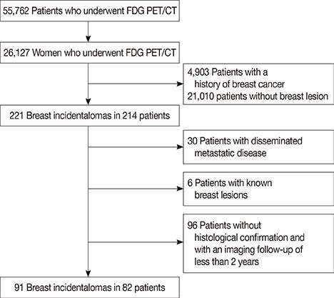
Figure 1 Consort diagram. There were 55,762 patients who underwent 18F-fluorodeoxyglucose (FDG) positron emission tomography with computed tomography (PET/CT) between March 2005 and October 2011. Data of 82 patients met the inclusion criteria and were used in this study.
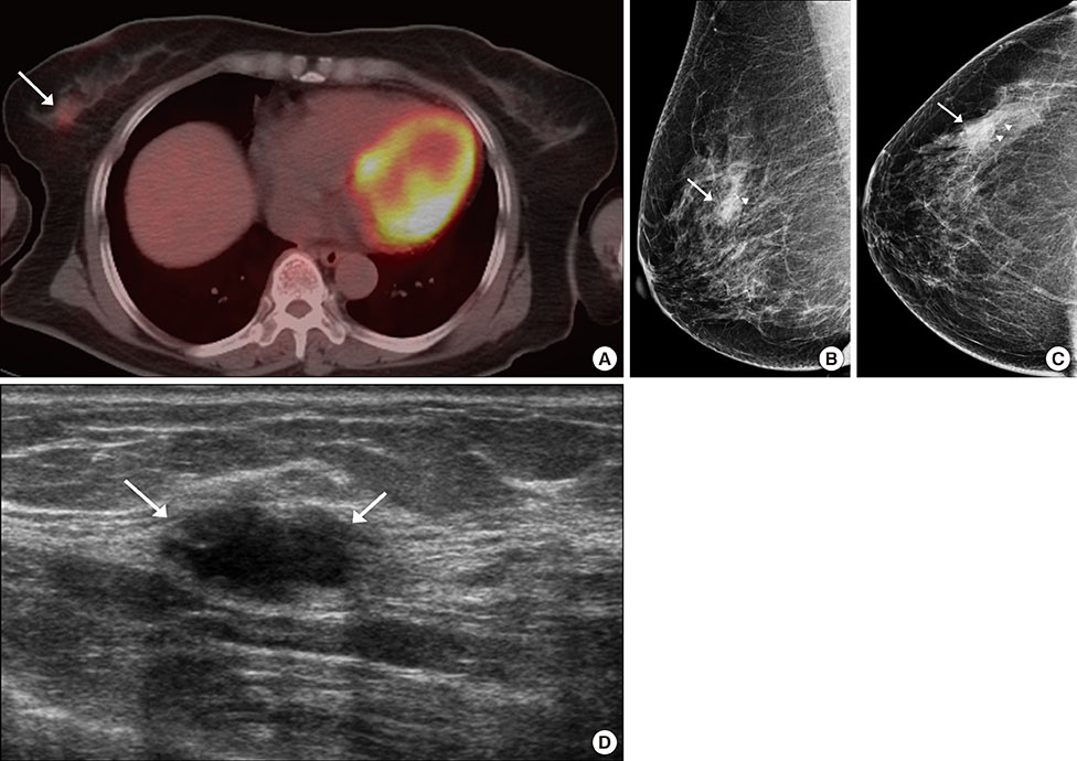
Figure 2 Breast incidentaloma in a 61-year-old woman with thyroid cancer. (A) Axial 18F-fluorodeoxyglucose (FDG) positron emission tomography with computed tomography (PET/CT) image shows focally increased uptake (maximum standardized uptake value, 2.6) (arrow) in the right upper outer breast. (B, C) Mammography shows a focal asymmetry (arrows) with suspicious microcalcifications (arrowheads). (D) Ultrasonography (US) image shows a 1.8-cm hypoechoic mass (arrows) with microlobulated margin and an oval shape in the right upper outer breast, correlating with the focal uptake on the PET/CT scan. Final assessment of the mass on US showed that it was of category 4c. The lesion was confirmed to be an invasive ductal carcinoma by USguided core biopsy.
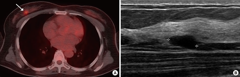
Figure 3 Breast incidentaloma in a 48-year-old woman with lymphoma. (A) Axial 18F-fluorodeoxyglucose (FDG) positron emission tomography with computed tomography (PET/CT) image shows focally increased uptake (maximum standardized uptake value, 5.7) (arrow) in the right upper outer breast. (B) Ultrasonography (US) image shows a 1.0-cm hypoechoic mass with circumscribed margin and an oval shape (asterisk) in the right upper outer breast, correlating with the focal uptake on the PET scan. Final assessment of the mass on US showed that it was of category 3. The lesion was confirmed to be an intraductal papilloma by excision.
Reference
-
1. Kostakoglu L, Agress H Jr, Goldsmith SJ. Clinical role of FDG PET in evaluation of cancer patients. Radiographics. 2003; 23:315–340.
Article2. Bohuslavizki KH, Klutmann S, Kröger S, Sonnemann U, Buchert R, Werner JA, et al. FDG PET detection of unknown primary tumors. J Nucl Med. 2000; 41:816–822.3. Bar-Shalom R, Valdivia AY, Blaufox MD. PET imaging in oncology. Semin Nucl Med. 2000; 30:150–185.
Article4. Beatty JS, Williams HT, Aldridge BA, Hughes MP, Vasudeva VS, Gucwa AL, et al. Incidental PET/CT findings in the cancer patient: how should they be managed? Surgery. 2009; 146:274–281.
Article5. Ishimori T, Patel PV, Wahl RL. Detection of unexpected additional primary malignancies with PET/CT. J Nucl Med. 2005; 46:752–757.6. Agress H Jr, Cooper BZ. Detection of clinically unexpected malignant and premalignant tumors with whole-body FDG PET: histopathologic comparison. Radiology. 2004; 230:417–422.
Article7. Korn RL, Yost AM, May CC, Kovalsky ER, Orth KM, Layton TA, et al. Unexpected focal hypermetabolic activity in the breast: significance in patients undergoing 18F-FDG PET/CT. AJR Am J Roentgenol. 2006; 187:81–85.
Article8. Chung A, Schoder H, Sampson M, Morrow M, Port E. Incidental breast lesions identified by 18F-fluorodeoxyglucose-positron emission tomography. Ann Surg Oncol. 2010; 17:2119–2125.
Article9. Kang BJ, Lee JH, Yoo IR, Kim SH, Choi JJ, Jeong SH, et al. Clinical significance of incidental finding of focal activity in the breast at 18F-FDG PET/CT. AJR Am J Roentgenol. 2011; 197:341–347.
Article10. Chae EY, Cha JH, Kim HH, Shin HJ, Kim HJ, Oh HY, et al. Analysis of incidental focal hypermetabolic uptake in the breast as detected by 18FFDG PET/CT: clinical significance and differential diagnosis. Acta Radiol. 2012; 53:530–535.
Article11. Kim MY, Cho N, Chang JM, Yun BL, Bae MS, Kang KW, et al. Mammography and ultrasonography evaluation of unexpected focal 18FFDG uptakes in breast on PET/CT. Acta Radiol. 2012; 53:249–254.
Article12. Dunne RM, O'Mahony D, Wilson G, McDermott R, O’Keeffe SA. The role of the breast radiologist in evaluation of breast incidentalomas detected on 18-fludeoxyglucose positron emission tomography/CT. Br J Radiol. 2013; 86:20130034.
Article13. Lim S, Lee EH, Park JM, Chang YW, Kim HH, Jeong SH. Role of combined BI-RADS assessment using mammography and sonography for evaluation of incidental hypermetabolic lesions in the breast on 18FFDG PET-CT. Acta Radiol. 2013; 54:1117–1124.
Article14. Mendelson EB, Bohn-Velez M, Berg WA, Whitman GJ, Feldman MI, Madjar H, et al. ACR BI-RADS ultrasound. American College of Radiology, BI-RADS Committee. ACR BI-RADS Atlas: Breast Imaging Reporting and Data System. Reston: American College of Radiology;2013.15. Kim EK, Ko KH, Oh KK, Kwak JY, You JK, Kim MJ, et al. Clinical application of the BI-RADS final assessment to breast sonography in conjunction with mammography. AJR Am J Roentgenol. 2008; 190:1209–1215.
Article16. Park VY, Kim MJ, Kim EK, Moon HJ. Second-look US: how to find breast lesions with a suspicious MR imaging appearance. Radiographics. 2013; 33:1361–1375.
Article17. Bertagna F, Treglia G, Orlando E, Dognini L, Giovanella L, Sadeghi R, et al. Prevalence and clinical significance of incidental F18-FDG breast uptake: a systematic review and meta-analysis. Jpn J Radiol. 2014; 32:59–68.
Article18. Litmanovich D, Gourevich K, Israel O, Gallimidi Z. Unexpected foci of 18F-FDG uptake in the breast detected by PET/CT: incidence and clinical significance. Eur J Nucl Med Mol Imaging. 2009; 36:1558–1564.
Article19. Beatty JS, Williams HT, Gucwa AL, Hughes MP, Vasudeva VS, Aldridge BA, et al. The predictive value of incidental PET/CT findings suspicious for breast cancer in women with non-breast malignancies. Am J Surg. 2009; 198:495–499.
Article20. Kumar R, Hawkins RA, Yeh BM, Wang ZJ. Focal fluorine-18 fluorodeoxyglucose-avid lesions without computed tomography correlate at whole-body positron emission tomography-computed tomography in oncology patients: how often are they malignant. Nucl Med Commun. 2011; 32:802–807.
Article21. Soo MS, Baker JA, Rosen EL. Sonographic detection and sonographically guided biopsy of breast microcalcifications. AJR Am J Roentgenol. 2003; 180:941–948.
Article22. Moon WK, Im JG, Koh YH, Noh DY, Park IA. US of mammographically detected clustered microcalcifications. Radiology. 2000; 217:849–854.
Article23. Lim HS, Yoon W, Chung TW, Kim JK, Park JG, Kang HK, et al. FDG PET/CT for the detection and evaluation of breast diseases: usefulness and limitations. Radiographics. 2007; 27:Suppl 1. S197–S213.
Article24. Berg WA, Blume JD, Cormack JB, Mendelson EB, Lehrer D, Böhm-Vélez M, et al. Combined screening with ultrasound and mammography vs mammography alone in women at elevated risk of breast cancer. JAMA. 2008; 299:2151–2163.
Article25. Avril N, Rosé CA, Schelling M, Dose J, Kuhn W, Bense S, et al. Breast imaging with positron emission tomography and fluorine-18 fluorodeoxyglucose: use and limitations. J Clin Oncol. 2000; 18:3495–3502.
Article26. Kumar R, Chauhan A, Zhuang H, Chandra P, Schnall M, Alavi A. Clinicopathologic factors associated with false negative FDG-PET in primary breast cancer. Breast Cancer Res Treat. 2006; 98:267–274.
Article27. Adejolu M, Huo L, Rohren E, Santiago L, Yang WT. False-positive lesions mimicking breast cancer on FDG PET and PET/CT. AJR Am J Roentgenol. 2012; 198:W304–W314.
Article28. Lee HJ, Kim EK, Kim MJ, Youk JH, Lee JY, Kang DR, et al. Observer variability of Breast Imaging Reporting and Data System (BI-RADS) for breast ultrasound. Eur J Radiol. 2008; 65:293–298.
Article29. Adibelli ZH, Ergenc R, Oztekin O, Ecevit S, Unal G, Abal Y. Observer variability of the Breast Imaging Reporting and Data System (BIRADS) lexicon for mammography. Breast Care (Basel). 2010; 5:11–16.
Article
- Full Text Links
-
- Actions
-
Cited
- CITED
-
- Close
- Share
-
- Similar articles
-
- Clinical Significance of Focal Breast Lesions Incidentally Identified by 18F-FDG PET/CT
- Hypermetabolic Axillary Mass on 18F FDG PET/CT: Breast Cancer Arising from Accessory Breast Tissue
- 18F-FDG PET/CT Findings in a Breast Cancer Patient with Concomitant Tuberculous Axillary Lymphadenitis
- Usefulness of 18F-fluoride PET/CT in Breast Cancer Patients with Osteosclerotic Bone Metastases
- 18F-FDG PET/CT and Sonographic Findings of Thyroid Incidentalomas
