A Case of Severe Strongyloidiasis in a Patient with Chronic Obstructive Pulmonary Disease Receiving Long-Term Steroid Therapy
- Affiliations
-
- 1Department of Internal Medicine and Laboratory Medicine, College of Medicine, Dongguk University, Gyongju, Korea. medione@dongguk.ac.kr
- 2Department of Parasitology, College of Medicine, Han-Yang University, Seoul, Korea.
- KMID: 2285052
- DOI: http://doi.org/10.3947/ic.2008.40.5.276
Abstract
- Strongyloides stercoralis is a soil-transmitted intestinal nematode that may cause long-lived auto-infection in the host. It is distributed worldwide, especially in the tropical and subtropical regions, but has been rarely reported in Korea. Chronic infections by S. stercoralis are mostly inapparent infections that carry nonspecific gastrointestinal and pulmonary symptoms. However, In immunocompromised patients such as those receiving long-term steroid therapy and patients with AIDS or malignant tumors, S. stercoralis can induce hyperinfection by autoinfection. This may lead to increased rate of complications such as resistance to chemotherapy and sepsis. In such cases mortality rate of up to 87% has been reported. We report a case of severe strongyloidiasis in a patient with chronic obstructive pulmonary disease who was receiving long-term steroid therapy. The chief complaint was repeated dyspnea and hematochezia, and strongyloidiasis was diagnosed by the presence of rhabditiform larvae of S. stercoralis in the fecal smear and isolation of filariform larvae from the stool culture. The patient developed septic shock during treatment with albendazole and showed clinical signs of hyperinfection of S. stercoralis. He eventually died despite aggressive treatment.
Keyword
MeSH Terms
Figure
-
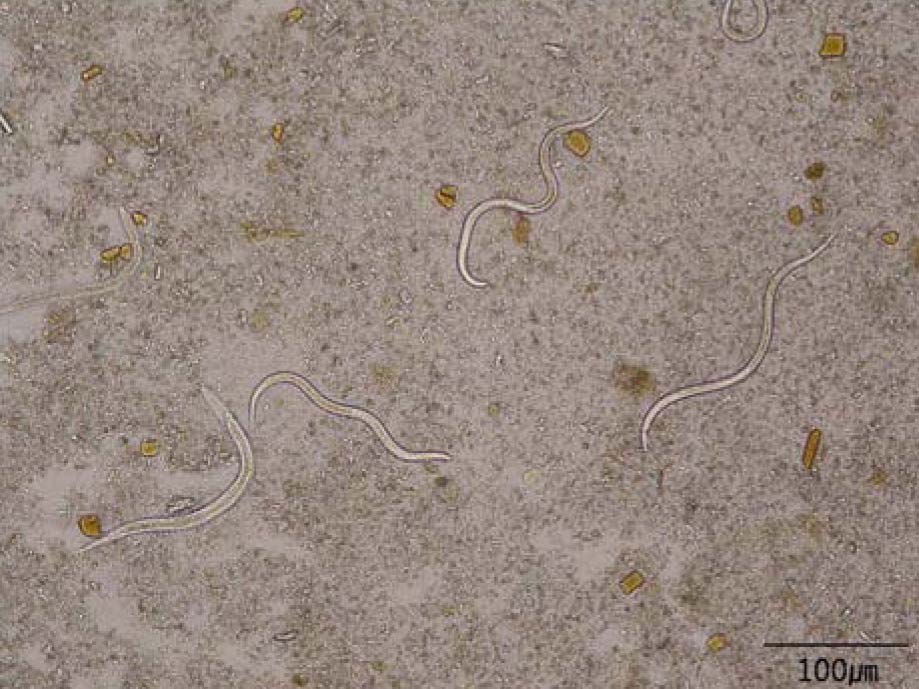
Figure 1 In patient's stool examination by the formalin-ether concentration method, several actively motile rhabditiform larvae were observed under the light microscope. scale bar=100 µm.
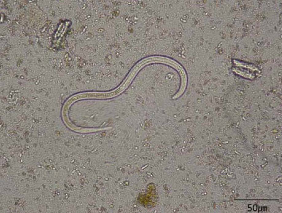
Figure 2 A rhabditiform larva of S. stercoralis, 251.4±5.8×20.6±0.6 µm in size. scale bar=50 µm.
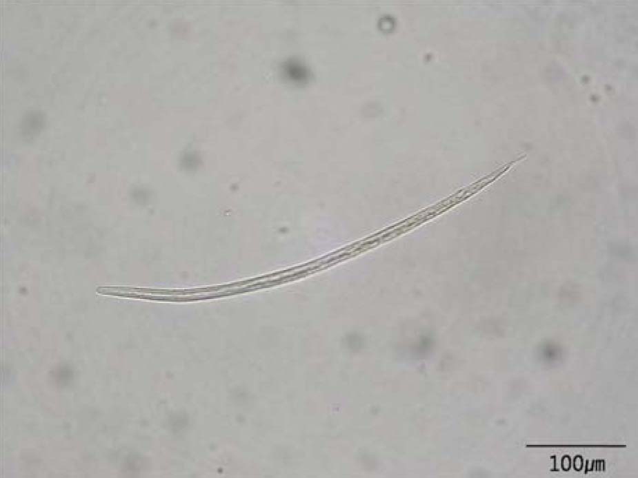
Figure 3 A filariform larva of S. stercolaris, 482.9±3.4×11.7±1.8 µm in size. scale bar=100 µm.
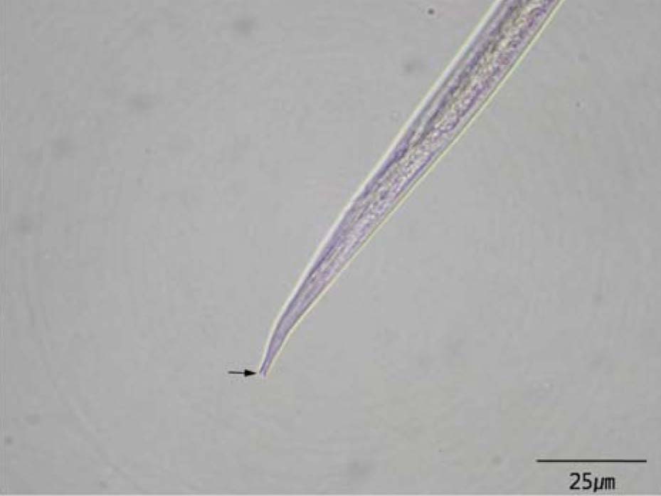
Figure 4 A filariform larva of S. stercolaris with characteristic notched tail. scale bar=25 µm.
Cited by 1 articles
-
Observation of the Free-living Adults of Strongyloides stercoralis from a Human Stool in Korea
Young-Hee Hong, Jong-Wan Kim, In-Soo Rheem, Jae-Soo Kim, Suk-Bae Kim, Jong-Yil Chai, Sang-Mee Guk, Seung-Ha Lee, Min Seo
Infect Chemother. 2009;41(2):105-108. doi: 10.3947/ic.2009.41.2.105.
Reference
-
1. Siddiqui AA, Berk SL. Diagnosis of Strongyloides stercoralis infection. Clin Infect Dis. 2001. 33:1040–1047.2. Lee SK, Shin BM, Khang SK, Chai JY, Kook J, Hong ST, Lee SH. Nine cases of strongyloidiasis in Korea. Korean J Parasitol. 1994. 32:49–52.
Article3. Kim SY, Kim NY, Lee KH, Gu MS, Chai JY, Kook J, Lee SH. A case of strongyloidiasis accompanied by duodenal ulcer. Korea J Parasit. 1992. 30:231–234.
Article4. Hong SJ, Shin JS, Kim SY. A case of strongloidiasis with hyperinfection syndrome. Korean J Parasitol. 1988. 26:221–226.
Article5. Lee SH, Ahn SJ, Koh IY, Jang JS, Park MA, Kim KH, Huh KY, Lee JH, Lee H, Han SY. A Case of Strongyloidiasis Associated with Intestinal obstruction in a Patient with Alcoholic Liver Disease. Infect Chemother. 2003. 35:467–470.6. Namisato S, Motomura K, Haranaga S, Hirata T, Toyama M, Shinzato T, Higa F, Saito A. Pulmonary strongyloidiasis in a patient receiving prednisolone therapy. Intern Med. 2004. 43:731–736.
Article7. Barr JR. Strongyloides stercoralis. Can Med Assoc J. 1978. 118:933–935.8. Ford J, Reiss-Levy E, Clark E, Dyson AJ, Schonell M. Pulmonary strongyloidiasis and lung abscess. Chest. 1981. 79:239–240.
Article9. Concha R, Harrington W Jr, Rogers AI. Intestinal strongyloidiasis: recognition, management, and determinants of outcome. J Clin Gastroenterol. 2005. 39:203–211.10. Fardet L, Gnreau T, Cabane J, Kettaneh A. Severe strongyloidiasis in corticosteroid-treated patients. Clin Microbiol Infect. 2006. 12:945–947.
Article11. Marcos LA, Terashima A, Dupont HL, Gotuzzo E. Strongyloides hyperinfection syndrome: an emerging global infectious disease. Trans R Soc Trop Med Hyg. 2008. 102:314–318.
Article12. Csermely L, Jaafar H, Kristensen J, Castella A, Gorka W, Chebli AA, Trab F, Alizadeh H, Hunyady B. Strongyloides hyper-infection causing life-threatening gastrointestinal bleeding. World J Gastroenterol. 2006. 12:6401–6404.
Article13. Zaha O, Hirata T, Kinjo F, Saito A. Strongyloidiasis-progress in diagnosis and treatment. Intern Med. 2000. 39:695–700.
Article14. Ghosh K, Ghosh K. Strongyloides stercoralis septicaemia following steroid therapy for eosinophilia: report of three cases. Trans R Soc Trop Med Hyg. 2007. 101:1163–1165.
Article15. Shikiya K, Kinjo N, Uehara T, Uechi H, Ohshiro J, Arakaki T, Kinjo F, Saito A, Iju M, Kobari K. Efficacy of ivermectin against Strongyloides stercoralis in humans. Intern Med. 1992. 31:310–312.
Article
- Full Text Links
-
- Actions
-
Cited
- CITED
-
- Close
- Share
-
- Similar articles
-
- Pulmonary Strongyloidiasis Masquerading as Exacerbation of Chronic Obstructive Pulmonary Disease
- Osteoporosis in Chronic Obstructive Pulmonary Disease
- Strongyloidiasis in a Diabetic Patient Accompanied by Gastrointestinal Stromal Tumor: Cause of Eosinophilia Unresponsive to Steroid Therapy
- A Case of Duodenal Ulcer Due to Coinfection with Strongyloides stericoralis and Cytomegalovirus
- Cor Pulmonale with Particular Reference to Chronic Obstructive Pulmonary Disease and Pulmonary Tuberculosis
