Aging Index using Photoplethysmography for a Healthcare Device: Comparison with Brachial-Ankle Pulse Wave Velocity
- Affiliations
-
- 1Division of Cardiology, Department of Internal Medicine, Hallym University College of Medicine, Chuncheon, Korea.
- 2Department of Electronics Engineering, College of Information and Electronics Engineering, Hallym University, Chuncheon, Korea. ajm@hallym.ac.kr
- KMID: 2284595
- DOI: http://doi.org/10.4258/hir.2015.21.1.30
Abstract
OBJECTIVES
Recent studies have emphasized the potential information embedded in peripheral fingertip photoplethysmogram (PPG) signals for the assessment of arterial wall stiffening during aging. For the discrimination of arterial stiffness with age, the brachial-ankle pulse wave velocity (baPWV) has been widely used in clinical applications. The second derivative of the PPG (acceleration photoplethysmogram [APG]) has been reported to correlate with the presence of atherosclerotic disorders. In this study, we investigated the association among age, the baPWV, and the APG and found a new aging index reflecting arterial stiffness for a healthcare device.
METHODS
The APG and the baPWV were simultaneously applied to assess the accuracy of the APG in measuring arterial stiffness in association with age. A preamplifier and motion artifact removal algorithm were newly developed to obtain a high quality PPG signal. In total, 168 subjects with a mean +/- SD age of 58.1 +/- 12.6 years were followed for two months to obtain a set of complete data using baPWV and APG analysis.
RESULTS
The baPWV and the B ratio of the APG indices were correlated significantly with age (r = 0.6685, p < 0.0001 and r = -0.4025, p < 0.0001, respectively). A regression analysis revealed that the c and d peaks were independent of age (r = -0.3553, p < 0.0001 and r = -0.3191, p < 0.0001, respectively).
CONCLUSIONS
We determined the B ratio, which represents an improved aging index and suggest that the APG may provide qualitatively similar information for arterial stiffness.
Keyword
MeSH Terms
Figure
-
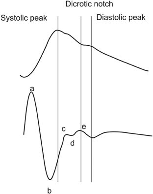
Figure 1 (Top) Original fingertip photoplethysmogram waveform. (Bottom) Second derivative of the photoplethysmogram (i.e., acceleration photoplethysmogram).
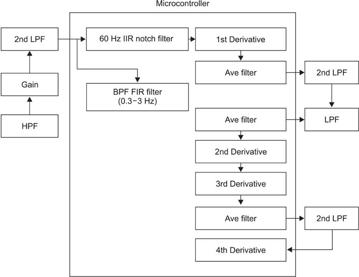
Figure 2 System configuration using microcontroller. 2nd LPF: second low-pass filter, HPF: high-pass filter, 60 Hz IIR notch filter: removal of 60 Hz power line with infinite impulse response, BPF FIR filter: band-pass filter finite impulse response filter, Ave filter: averaging filter, 1st, 2nd, 3rd, and 4th derivative: the order of the derivative of the photoplethysmogram waveform.
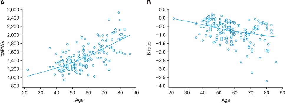
Figure 3 Scatter diagrams of the relationship between the brachial-ankle pulse wave velocity (baPWV) and age (A) and the relationship between the B ratio of the acceleration photoplethysmogram and age (B).
Cited by 2 articles
-
Wave Detection in Acceleration Plethysmogram
Jae Mok Ahn
Healthc Inform Res. 2015;21(2):111-117. doi: 10.4258/hir.2015.21.2.111.New Aging Index Using Signal Features of Both Photoplethysmograms and Acceleration Plethysmograms
Jae Mok Ahn
Healthc Inform Res. 2017;23(1):53-59. doi: 10.4258/hir.2017.23.1.53.
Reference
-
1. Nitzan M, Babchenko A, Khanokh B, Landau D. The variability of the photoplethysmographic signal: a potential method for the evaluation of the autonomic nervous system. Physiol Meas. 1998; 19(1):93–102.
Article2. Huotari M, Vehkaoja A, Maatta K, Kostamovaara J. Photoplethysmography and its detailed pulse waveform analysis for arterial stiffness. J Struct Mech. 2011; 44(4):345–362.3. Elgendi M. On the analysis of fingertip photoplethysmogram signals. Curr Cardiol Rev. 2012; 8(1):14–25.
Article4. Yousef Q, Reaz MB, Ali MA. The analysis of PPG morphology: investigating the effects of aging on arterial compliance. Meas Sci Rev. 2012; 12(6):266–271.
Article5. Sugawara J, Hayashi K, Yokoi T, Cortez-Cooper MY, DeVan AE, Anton MA, et al. Brachial-ankle pulse wave velocity: an index of central arterial stiffness? J Hum Hypertens. 2005; 19(5):401–406.
Article6. Kang S, Fan HM, Li J, Fan LY, Miao AY, Bao Y, et al. Relationship of arterial stiffness and early mild diastolic heart failure in general middle and aged population. Eur Heart J. 2010; 31(22):2799–2807.
Article7. Yamashina A, Tomiyama H, Takeda K, Tsuda H, Arai T, Hirose K, et al. Validity, reproducibility, and clinical significance of noninvasive brachial-ankle pulse wave velocity measurement. Hypertens Res. 2002; 25(3):359–364.
Article8. Tomiyama H, Yamashina A, Arai T, Hirose K, Koji Y, Chikamori T, et al. Influences of age and gender on results of noninvasive brachial-ankle pulse wave velocity measurement: a survey of 12517 subjects. Atherosclerosis. 2003; 166(2):303–309.
Article9. Blazek R, Lee C. Multi-resolution linear model comparison for detection of dicrotic notch and peak in blood volume pulse signals. Anal Biomed Signals Images. 2010; 20:378–386.
- Full Text Links
-
- Actions
-
Cited
- CITED
-
- Close
- Share
-
- Similar articles
-
- Usefulness of ankle brachial pressure index measured using photoplethysmography and automated blood pressure measurement device
- Pulse wave velocity and ankle brachial index in normal adolescents
- Usefulness of Brachial-Ankle Pulse Wave Velocity and Ankle-Brachial Index as Predictors of Early Age-Related Maculopathy
- Association between Aging and Changes in the Ankle-Brachial Index after Exercise in Patients with Chest Pain
- Relationship between Cardiac Autonomic Neuropathy and Brachial-ankle Pulse Wave Velocity in Type 2 Diabetics
