GFP-tagged E. coli shows bacterial distribution in mouse organs: pathogen tracking using fluorescence signal
- Affiliations
-
- 1Division of High-Risk Pathogen Research, Center for Infectious Diseases, Korea National Institute of Health, Cheongwon, Korea. khong@nih.go.kr
- 2Laboratory of Molecular Imaging and Therapy, Cancer Research Institute, Seoul National University College of Medicine, Seoul, Korea.
- 3Department of Nuclear Medicine, Cancer Imaging Center, Seoul National University Hospital, Seoul, Korea.
- KMID: 2278792
- DOI: http://doi.org/10.7774/cevr.2012.1.1.83
Abstract
- PURPOSE
In vaccine efficacy evaluation, visualization of pathogens in whole organism at each time point would be able to reduce the consuming animals and provide the in vivo information within consistent background with identical organism.
MATERIALS AND METHODS
Using IVIS spectrum whole live-animal imaging system, fluorescent intensity was optimized and visualized proportionately by concentrating Escherichia coli MC1061 strain which expresses GFP (E. coli-GFP) in BALB/C mice after injection.
RESULTS
Local distribution of disseminated E. coli-GFP was traced in each organ by fluorescence. Detached organ showed more obvious fluorescent signal, and intestine showed strongest fluorescent signal.
CONCLUSION
This in vivo imaging method using GFP-tagged pathogen strain suggest quantified infected pathogens by fluorescence intensity in whole animals can provide the information about the localization and distribution after infection.
MeSH Terms
Figure
-
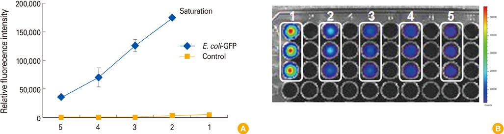
Fig. 1 Fluorescent signal of E. coli-GFP strain. (A) Relative fluorescence intensity of E. coli-GFP. Fluorescent signal intensity was increased by bacteria count. Intensity of highly concentrated bacteria (No. 1) appeared as saturated signal. Bacteria numbers matched with each number (1-5) are presented at Table 1. (B) Visualization of serial diluted E. coli-GFP. In pseudo-color covered image, the high intensity of fluorescence appeared as red color and low intensity of fluorescence as blue. E. coli-GFP, Escherichia coli MC1061 strain which expresses green fluorescent protein.
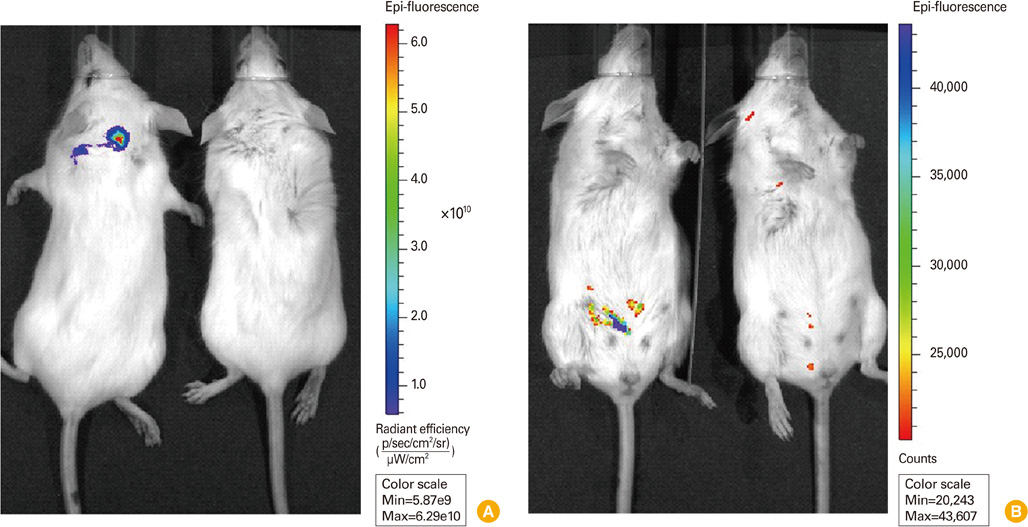
Fig. 2 Visualization of E. coli-GFP strain in mice by in vivo imaging. (A) Fluorescent imaging of E. coli-GFP injected with intradermal route. 5×10⁹ CFU (500 µL) of E. coli-GFP was subcutaneously injected and dorsal side of mice were analyzed. Left animal, injected mouse; right animal, uninjected mouse; pseudo-color, red (high) to blue (low). (B) Fluorescent imaging of E. coli-GFP injected with intraperitoneal route. 3.5×10⁹ CFU (350 µL) of E. coli-GFP was injected to abdominal cavity and ventral side of injected mice were analyzed. Left animal, injected mouse; right animal, uninjected mouse; pseudo-color, blue (high) to red (low). E. coli-GFP, Escherichia coli MC1061 strain which expresses green fluorescent protein; CFU, colony-forming unit.
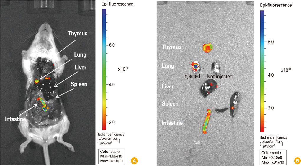
Fig. 3 Visualization of E. coli-GFP strain in mice by anatomical imaging. (A) Fluorescent imaging of E. coli-GFP injected to each organ. Mouse was administered euthanasia and opened the chest and abdominal cavity for direct injection of bacteria. 0.5-1.0×10⁹ CFU of E. coli-GFP was injected to each organ. (B) Fluorescent imaging of detached organ of (A). Left lung was not injected with bacteria. Pseudo-color, blue (high) to red (low). E. coli-GFP, Escherichia coli MC1061 strain which expresses green fluorescent protein; CFU, colony-forming unit.
Reference
-
1. Golovliov I, Baranov V, Krocova Z, Kovarova H, Sjostedt A. An attenuated strain of the facultative intracellular bacterium Francisella tularensis can escape the phagosome of monocytic cells. Infect Immun. 2003. 71:5940–5950.
Article2. Doyle TC, Burns SM, Contag CH. In vivo bioluminescence imaging for integrated studies of infection. Cell Microbiol. 2004. 6:303–317.
Article3. Shen H, Harris G, Chen W, Sjostedt A, Ryden P, Conlan W. Molecular immune responses to aerosol challenge with Francisella tularensis in mice inoculated with live vaccine candidates of varying efficacy. PLoS One. 2010. 5:e13349.
Article4. Nham T, Filali S, Danne C, Derbise A, Carniel E. Imaging of bubonic plague dynamics by in vivo tracking of bioluminescent Yersinia pestis. PLoS One. 2012. 7:e34714.
Article5. Sanz P, Teel LD, Alem F, Carvalho HM, Darnell SC, O'Brien AD. Detection of Bacillus anthracis spore germination in vivo by bioluminescence imaging. Infect Immun. 2008. 76:1036–1047.
Article6. Green M, Choules G, Rogers D, Titball RW. Efficacy of the live attenuated Francisella tularensis vaccine (LVS) in a murine model of disease. Vaccine. 2005. 23:2680–2686.
Article7. Bina XR, Miller MA, Bina JE. Construction of a bioluminescence reporter plasmid for Francisella tularensis. Plasmid. 2010. 64:156–161.
Article8. Miller MA, Stabenow JM, Parvathareddy J, et al. Visualization of murine intranasal dosing efficiency using luminescent Francisella tularensis: effect of instillation volume and form of anesthesia. PLoS One. 2012. 7:e31359.
Article9. Contag CH, Bachmann MH. Advances in in vivo bioluminescence imaging of gene expression. Annu Rev Biomed Eng. 2002. 4:235–260.
Article10. Shaner NC, Steinbach PA, Tsien RY. A guide to choosing fluorescent proteins. Nat Methods. 2005. 2:905–909.
Article11. Youn H, Hong KJ. In vivo noninvasive small animal molecular imaging. Osong Public Health Res Perspect. 2012. 3:48–59.12. Kawasaki ES, Player A. Nanotechnology, nanomedicine, and the development of new, effective therapies for cancer. Nanomedicine. 2005. 1:101–109.
Article13. Pulendran B, Ahmed R. Translating innate immunity into immunological memory: implications for vaccine development. Cell. 2006. 124:849–863.
Article14. Pechous RD, McCarthy TR, Zahrt TC. Working toward the future: insights into Francisella tularensis pathogenesis and vaccine development. Microbiol Mol Biol Rev. 2009. 73:684–711.
Article15. Hagenaars N, Mania M, de Jong P, et al. Role of trimethylated chitosan (TMC) in nasal residence time, local distribution and toxicity of an intranasal influenza vaccine. J Control Release. 2010. 144:17–24.
Article
- Full Text Links
-
- Actions
-
Cited
- CITED
-
- Close
- Share
-
- Similar articles
-
- Unfolded Histidine-Tagged Protein is Immobilized to Nitrilotriacetic Acid-Nickel Beads, But Not the Nickel-Coated Glass Slide
- Development of dual reporter imaging system for Francisella tularensis to monitor the spatio-temporal pathogenesis and vaccine efficacy
- Expression of Green Fluorescent Protein in Both Spodoptera frugiperda Cells and Bombyx mori Larvae by Ac-Bm Hybrid Virus
- Tracking the Fate of Muscle-derived Stem Cells: an Insight into the Distribution and Mode of Action
- In vitro MRI and Characterization of Rat Mesenchymal Stem Cells Transduced with Ferritin as MR Reporter Gene
