Successful Treatment of Rectus Femoris Calcification with Ultrasound-guided Injection: A Case Report
- Affiliations
-
- 1Department of Rheumatology, Presbyterian Medical Center, Jeonju, Korea.
- 2Department of Anesthesiology and Pain Medicine, Wonkwang University Hospital, School of Medicine, Iksan, Korea. kydpain@hanmail.net
- 3Institute of Wonkwang Medical Science, Iksan, Korea.
- 4Department of Anesthesiology and Pain Medicine, Na-eun Hospital, Iksan, Korea.
- KMID: 2278253
- DOI: http://doi.org/10.3344/kjp.2015.28.1.52
Abstract
- Painful periarticular calcification most commonly occurs within the rotator cuff of the shoulder and rarely around the elbow, hip, foot, and neck. As acute inflammatory reaction develops, severe pain, exquisite tenderness, local swelling, and limitation of motion with pain occur. In case of calcific tendinitis of the shoulder, it can be easily diagnosed according to the symptoms and with x-ray. However, in lesions of the hip, as it is a rare location and usually involves pain in the posterolateral aspect of the thigh, which can simulate radicular pain from a lumbar intervertebral disc, it could be difficult to diagnose. Hence, physicians usually focus on lumbar lesions; therefore, misdiagnosis is common and leads to a delayed management. Here, we report the case of a 30-year-old female patient with calcific tendinitis of the rectus femoris that was successfully managed with ultrasound-guided steroid injection. This study offers knowledge about the rectus femoris calcification.
MeSH Terms
Figure
-
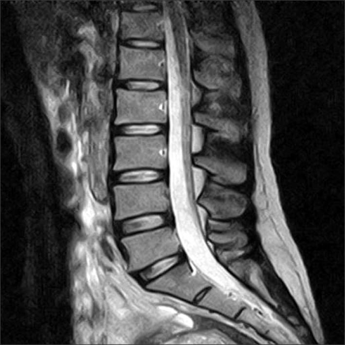
Fig. 1 Magnetic resonance imaging finding of the lumbar spine, sagittal plane.

Fig. 2 Plain hip AP showed a small amorphous calcification near the right anterior inferior iliac spine, which is the attachment site of the rectus tendon, suggesting calcific tendinitis of rectus femoris (A). The lesion was more clearly defined when the patient posed frog leg position (B).

Fig. 3 Ultrasound scanning over the hip area showing calcification around the rectus femoris with mild fluid collection. AIIS: anterior inferior iliac spine, Ca: calcification, Rf: rectus femoris.
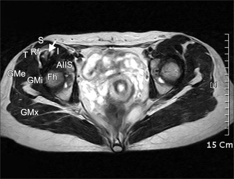
Fig. 4 Magnetic resonance imaging finding of the hip, axial plane. Calcification was noted in the right rectus femoris tendon showed thickening with small amount of fluid collection at the intermuscular fat plane. White arrow indicates calcification. AIIS: Anterior inferior iliac spine, Rf: Rectus femoris muscle and tendon, S: Sartorius muscle, I: Iliopsoas muscle, T: Tensor fasciae latae muscle, GMx: Gluteus maximus, GMe: Gluteus medius, GMi: Gluteus minimus.
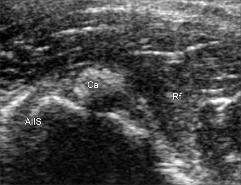
Fig. 5 Ultrasonography scanning over the hip area, after 6 weeks. Decreased echogenicity of surrounding fat with decreased amount of fluid collection and size was observed.
Reference
-
1. King JW, Vanderpool DW. Calcific tendonitis of the rectus femoris. Am J Orthop. 1967; 9:110–111. PMID: 6046364.2. Sarkar JS, Haddad FS, Crean SV, Brooks P. Acute calcific tendinitis of the rectus femoris. J Bone Joint Surg Br. 1996; 78:814–816. PMID: 8836078.
Article3. Yun HH, Park JH, Park JW, Lee JW. Calcific tendinitis of the rectus femoris. Orthopedics. 2009; 32:490. PMID: 19634853.
Article4. Hajiroussou VJ, Webley M. Familial calcific periarthritis. Ann Rheum Dis. 1983; 42:469–470. PMID: 6882045.
Article5. Holt PD, Keats TE. Calcific tendinitis: a review of the usual and unusual. Skeletal Radiol. 1993; 22:1–9. PMID: 8430339.
Article6. Cannon RB, Schmid FR. Calcific periarthritis involving multiple sites in identical twins. Arthritis Rheum. 1973; 16:393–396. PMID: 4708018.
Article7. Uhthoff HK, Sarkar K, Maynard JA. Calcifying tendinitis: a new concept of its pathogenesis. Clin Orthop Relat Res. 1976; 164–168. PMID: 954272.8. Sakai T, Shimaoka Y, Sugimoto M, Koizumi T. Acute calcific tendinitis of the gluteus medius: a case report with serial magnetic resonance imaging findings. J Orthop Sci. 2004; 9:404–407. PMID: 15278780.
Article9. Goldenberg RR, Leventhal GS. Supratrochanteric calcification. J Bone Joint Surg Am. 1936; 18:205–211.10. Lecocq E. Peritrochanteric bursitis: report of a case. J Bone Joint Surg Am. 1931; 13:872–873.11. Chow HY, Recht MP, Schils J, Calabrese LH. Acute calcific tendinitis of the hip: case report with magnetic resonance imaging findings. Arthritis Rheum. 1997; 40:974–977. PMID: 9153562.
Article
- Full Text Links
-
- Actions
-
Cited
- CITED
-
- Close
- Share
-
- Similar articles
-
- Calcific Tendinitis of the Rectus Femoris Around the Hip Joint
- Successful Treatment of Abdominal Cutaneous Entrapment Syndrome Using Ultrasound Guided Injection
- Usefulness of Ultrasound for Carpal Tunnel Syndrome Proven in Meta-Analysis Studies
- Calcific Tendinitis of the Rectus Femoris: A Case Report
- Accuracy of Needle Placement in Cadavers: Non-Guided Versus Ultrasound-Guided
