A Case of Papillary Thyroid Cancer Recurring as an Esophageal Submucosal Tumor
- Affiliations
-
- 1Department of Internal Medicine, Pusan National University School of Medicine, Busan, Korea. doc0224@chol.com
- KMID: 2274884
- DOI: http://doi.org/10.4068/cmj.2012.48.1.60
Abstract
- A 75-year-old woman who underwent a total thyroidectomy for papillary thyroid cancer 7 years previously presented with a palpable neck mass. Computed tomography (CT) showed two metastatic masses on the thyroid bed and another mass that looked benign originating from the esophageal wall. Endoscopic ultrasonography (EUS) showed a hypoechoic mass in the esophageal wall that looked similar to a gastrointestinal stromal tumor. The mass on the esophagus had intense fluorodeoxyglucose (FDG) uptake in positron emission tomography-computed tomography (PET-CT), which suggested the possibility of malignancy. Subsequently, after surgery, the mass in the esophagus was confirmed as a metastasis from the thyroid papillary carcinoma. Here we report this unusual case of papillary thyroid cancer that recurred as an esophageal submucosal tumor.
Keyword
MeSH Terms
Figure
-
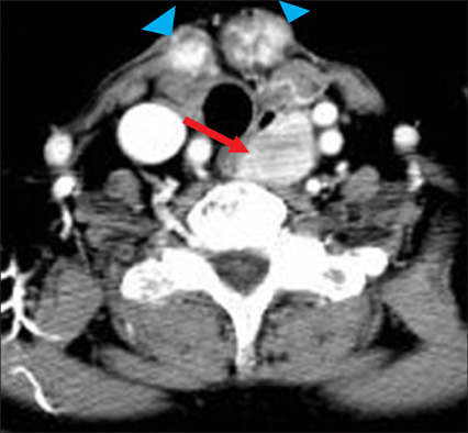
FIG. 1 Computed tomography findings. Two well-enhanced ill-defined masses (blue arrow head) are seen on the anterior aspect of the thyroid bed. An approximately 24-mm sized well-defined mass (red arrow) is located on the left side of the esophagus.

FIG. 2 Endosonography findings. (A) A round mass covered with normal overlying mucosa is seen in the upper esophagus 3 cm distal to the upper esophageal sphincter. (B, C) A relatively poorly demarcated, 2.7×2.4 cm sized hypoechoic nonhomogeneous mass originates from the third (submucosal) and fourth (muscularis propria) layer.
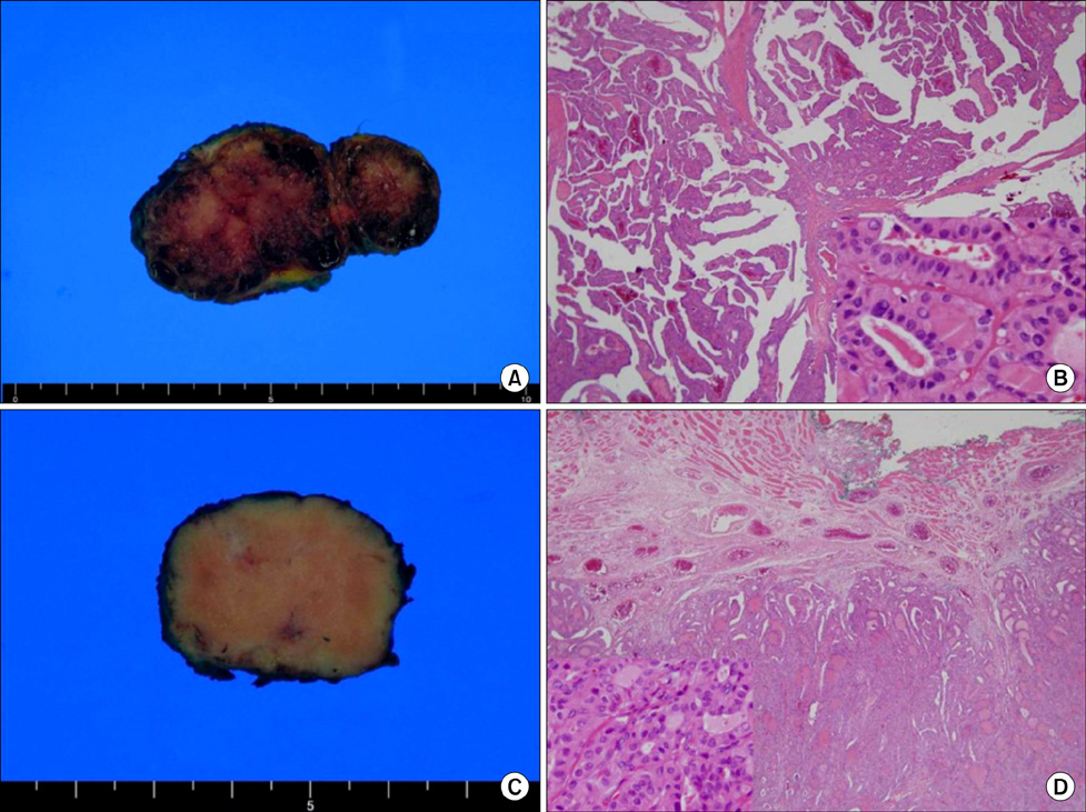
FIG. 3 Gross and microscopic findings. (A, B) Two metastatic masses on the thyroid beds. Two masses with fibrous capsule are seen. The cut surface is papillary with multiple foci of cystic change. Microscopically, the tumor cells exhibited predominantly papillary structures (H&E stain, ×40). Inset: Nuclei of tumor cells have typical intranuclear inclusions and groovings, which are the typical characteristics of papillary cancer cells (H&E stain, ×400). (C, D) The cut surface of the esophageal mass is light brown in color with focal fibrosis and hemorrhage. Microscopically, the tumor of the esophageal mass is located in the submucosal and muscularis propria layer. The tumor cells also showed a papillary pattern in appearance (H&E stain, ×40). Inset: Nuclei of tumor cells also have intranuclear inclusions and groovings, which are typical characteristics of papillary cancer cells (H&E stain, ×400).
Cited by 1 articles
-
Submucosal Esophageal Metastasis in a Patient with Poorly Differentiated Thyroid Carcinoma: a Case Report
Mi Jin Kim, Cheol Seung Kim, Young Sam Park, Eun Hye Choi, Kyu Dam Han
J Endocr Surg. 2017;17(3):131-137. doi: 10.16956/jes.2017.17.3.131.
Reference
-
1. Agha FP. Secondary neoplasms of the esophagus. Gastrointest Radiol. 1987. 12:187–193.
Article2. Hoie J, Stenwig AE, Kullmann G, Lindegaard M. Distant metastases in papillary thyroid cancer. A review of 91 patients. Cancer. 1988. 61:1–6.3. Lupoli GA, Fonderico F, Colarusso S, Panico A, Cavallo A, Di Micco L, et al. Current management of differentiated thyroid carcinoma. Med Sci Monit. 2005. 11:RA368–RA373.4. McCaffrey JC. Aerodigestive tract invasion by well-differentiated thyroid carcinoma: diagnosis, management, prognosis, and biology. Laryngoscope. 2006. 116:1–11.
Article5. McCaffrey JC. Evaluation and treatment of aerodigestive tract invasion by well-differentiated thyroid carcinoma. Cancer Control. 2000. 7:246–252.
Article6. Ohshima A, Yamashita H, Noguchi S. Endoscopic ultrasonography in the evaluation of thyroid cancer invasion into the esophagus. Surgery. 2000. 127:478–479.
Article7. Lee MY, Kim SE, Kim HC, Han SH, Shin DH, Kim DH, et al. Direct invasion of thyroid papillary carcinoma to esophagus presenting as an intraluminal polypoid mass which causes hematemesis. Korean J Gastrointest Endosc. 2001. 23:466–469.8. Cho DS, Ahn BY, Oh HT, Lee DS, Han DH, Kim SY, et al. Case of occult papillary carcinoma of thyroid, invaded trachea and esophagus. Tuberc Respir Dis. 1997. 44:1125–1131.
Article9. Kang HS, Park SH, Kim DJ, Won TS, Cho SJ, Lee TU. A case of locally invasive thyroid papillary cancer diagnosed by esophagoscopy. Korean J Gastrointest Endosc. 2009. 38:339–342.
- Full Text Links
-
- Actions
-
Cited
- CITED
-
- Close
- Share
-
- Similar articles
-
- Endoscopic Submucosal Dissection in the Treatment of Patients With Papillary Early Gastric Cancer
- Submucosal Esophageal Metastasis in a Patient with Poorly Differentiated Thyroid Carcinoma: a Case Report
- A Case of Cystic Lymph Node Metastasis from Thyroid Papillary Microcarcinoma
- Endoscopic Submucosal Dissection for Esophageal Squamous Cell Carcinoma
- A Case of Trophoblastic Tumor Associated with Papillary Thyroid Cancer
