Surface roughness analysis of ceramic bracket slots using atomic force microscope
- Affiliations
-
- 1Department of Orthodontics, College of Dentistry, Kyunghee University, Seoul, Korea.
- 2Department of Biomedical Engineering, College of Medicine, Kyunghee University, Seoul, Korea.
- 3Department of Orthodontics, College of Dentistry, Kyunghee University, Seoul, Korea. ygpark@khu.ac.kr
- KMID: 2273220
- DOI: http://doi.org/10.4041/kjod.2010.40.5.294
Abstract
OBJECTIVE
This study was designed to measure the surface roughness at the slot floor of various ceramic brackets.
METHODS
One kind of stainless steel bracket (Succes(R)), two kinds of monocrystalline brackets (Inspire Ice(R), Perfect(R)) and two kinds of polycrystalline brackets (Crystalline V(R), Invu(R)) were examined. Atomic force microscopy (AFM) was used to measure the surface roughness of each bracket. Data acquisition and processing were performed using SPIP(TM).
RESULTS
The differences in values of Sa, Sq, and Sz in Invu(R) and Inspire Ice(R) were not statistically different from the control group Succes(R). The values of Sa, Sq, and Sz of Perfect(R) and Crystalline V(R) were greater than those of Succes(R). Differences of all the Sa, Sq, and Sz values between Perfect(R) and Crystalline V(R) were not statistically significant.
CONCLUSIONS
It is concluded that the slot surfaces of Succes(R), Inspire Ice(R), and Invu(R) were smooth compared to those of Crystalline V(R) and Perfect(R).
Keyword
MeSH Terms
Figure
-
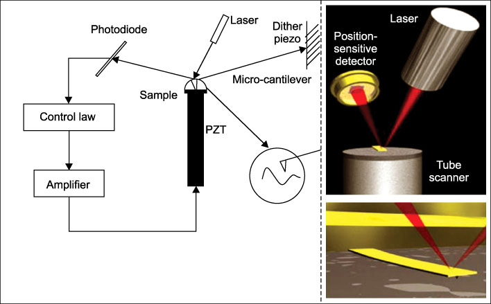
Fig. 1 A schematic view of atomic force microscopy imaging systems.20
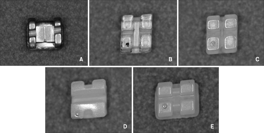
Fig. 2 Control group (A) and experimental groups (B - E). A, Succes® (Tomy); B, Inspire Ice® (Ormco); C, Perfect® (Hubit); D, Crystalline V® (Tomy); E, Invu® (TP Orthodontics).
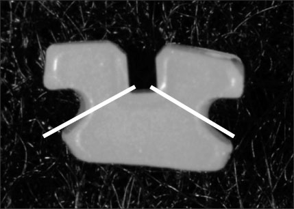
Fig. 3 Direction of grinding. Bracket wings were grinded to open slot base.

Fig. 4 Atomic force microscope (Nanostation II™).

Fig. 5 Optical microscopic images of several brackets (×500). A, Succes®; B, Inspire Ice®; C, Perfect®; D, Crystalline V®; E, Invu®.
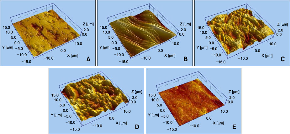
Fig. 6 Three-dimensional topographic images of control group (A) and experimental groups (B - E). A, Succes®; B, Inspire Ice®; C, Perfect®; D, Crystalline V®; E, Invu®.
Cited by 1 articles
-
Surface analysis of metal clips of ceramic self-ligating brackets
Kyung Sook Kim, Se Jik Han, Tae-Hee Lee, Tae-Joon Park, Samjin Choi, Yoon-Goo Kang, Ki-Ho Park
Korean J Orthod. 2019;49(1):12-20. doi: 10.4041/kjod.2019.49.1.12.
Reference
-
1. Angolkar PV, Kapila S, Duncanson MG Jr, Nanda RS. Evaluation of friction between ceramic brackets and orthodontic wires of four alloys. Am J Orthod Dentofacial Orthop. 1990. 98:499–506.
Article2. Pratten DH, Popli K, Germane N, Gunsolley JC. Frictional resistance of ceramic and stainless steel orthodontic brackets. Am J Orthod Dentofacial Orthop. 1990. 98:398–403.
Article3. Sung HM, Park YC. Comparison of the frictional resistance between orthodontic bracket & archwire. Korean J Orthod. 1991. 21:543–559.4. Riley JL, Garrett SG, Moon PC. Frictional forces of ligated plastic and metal edgewise brackets. J Dent Res. 1979. 58:A21.5. Drescher D, Bourauel C, Schumacher HA. Frictional forces between bracket and arch wire. Am J Orthod Dentofacial Orthop. 1989. 96:397–404.
Article6. Kusy RP, Whitley JQ, Mayhew MJ, Buckthal JE. Surface roughness of orthodontic archwires via laser spectroscopy. Angle Orthod. 1988. 58:33–45.7. Sims AP, Waters NE, Birnie DJ, Pethybridge RJ. A comparison of the forces required to produce tooth movement in vitro using two self-ligating brackets and a pre-adjusted bracket employing two types of ligation. Eur J Orthod. 1993. 15:377–385.
Article8. Omana HM, Moor RN, Bagby MD. Frictional properties of metal and ceramic brackets. J Clin Orthod. 1992. 26:425–432.9. Dowling PA, Jones WB, Lagerstrom L, Sandham JA. An investigation into the behavioral characteristics of orthodontic elastomeric modules. Br J Orthod. 1998. 25:197–202.
Article10. Bednar JR, Gruendeman GW, Sandrik JL. A comparative study of frictional forces between orthodontic brackets and arch wires. Am J Orthod Dentofacial Orthop. 1991. 100:513–522.
Article11. Saunders CR, Kusy RP. Surface topography and frictional characteristics of ceramic brackets. Am J Orthod Dentofacial Orthop. 1994. 106:76–87.
Article12. Zinelis S, Eliades T, Eliades G, Makou M, Silikas N. Comparative assessment of the roughness, hardness, and wear resistance of aesthetic bracket materials. Dent Mater. 2005. 21:890–894.
Article13. Horton CB, Paulus HM, Pelleu GB, Rudolph JJ. An evaluation of commercial pastes for finishing composite resin surfaces. J Prosthet Dent. 1977. 37:674–679.
Article14. Roulet JF, Roulet-Mehrens TK. The surface roughness of restorative materials and dental tissues after polishing with prophylaxis and polishing pastes. J Periodontol. 1982. 53:257–266.
Article15. Cassinelli C, Morra M. Atomic force microscopy studies of the interaction of a dentin adhesive with tooth hard tissue. J Biomed Mater Res. 1994. 28:1427–1431.
Article16. Marshall GW Jr, Balooch M, Tench RJ, Kinney JH, Marshall SJ. Atomic force microscopy of acid effects on dentin. Dent Mater. 1993. 9:265–268.
Article17. Marshall GW Jr, Balooch M, Kinney JH, Marshall SJ. Atomic force microscopy of conditioning agents on dentin. J Biomed Mater Res. 1995. 29:1381–1387.
Article18. Park HK. Atomic microscopy in kidney research. The 5th Congress of nephrology. 2007. 11. Kyunghee Univ;127–132.19. Park WQ. A study on surface roughness of metals after finishing and polishing - an atomic force microscope study. 1998. A master's thesis Kyunghee Univ.20. Jalili N, Laxminarayana K. A review of atomic force microscopy imaging systems: application to molecular metrology and biological sciences. Mechatronics. 2004. 14:907–945.
Article21. Widu F, Drescher D, Junker R, Bourauel C. Corrosion and biocompatibility of orthodontic wires. J Mater Sci Mater Med. 1999. 10:275–281.22. Huang HH. Variation in surface topography of different NiTi orthodontic archwires in various commercial fluoride-containing environments. Dent Mater. 2007. 23:24–33.
Article23. Bourauel C, Fries T, Drescher D, Plietsch R. Surface roughness of orthodontic wires via atomic force microscopy, laser specular reflectance, and profilometry. Eur J Orthod. 1998. 20:79–92.
Article24. Lin MC, Lin SC, Lee TH, Huang HH. Surface analysis and corrosion resistance of different stainless steel orthodontic brackets in artificial saliva. Angle Orthod. 2006. 76:322–329.25. Kim HS, Woo JH. A study on surface roughness of composite resins after finishing and polishing - an atomic force microscope study. J Korean Acad Prosthodont. 1997. 35:719–741.26. Valois CR, Silva LP, Azebedo RB. Atomic force microscopy study of stainless-steel and nickel-titanium files. J Endod. 2005. 31:882–885.
Article27. Lamolle SF, Monjo M, Lyngstadaas SP, Ellingsen JE, Haugen HJ. Titanium implant surface modification by cathodic reduction in hydrofluoric acid: surface characterization and in vivo performance. J Biomed Mater Res A. 2009. 88:581–588.28. El Feninat F, Ellis TH, Sacher E, Stangel I. A tapping mode AFM study of collapse and denaturation in dentinal collagen. Dent Mater. 2001. 17:284–288.
Article29. Freitas A, Espejo LC, Botta SB, Teixeira FS, Luz MA, Garone-Netto N, et al. AFM analysis of bleaching effects on dental enamel microtopography. Appl Surf Sci. 2010. 256:2915–2919.
Article30. Graber TM. Orthodontics: current principles and techniques. 2000. 3th ed. St. Louis: Elsevier;330–332.31. Birnie D. Ceramic brackets. Br J Orthod. 1990. 17:71–75.
Article32. Fox NA, McCabe JF. An easily removable ceramic bracket? Br J Orthod. 1992. 19:305–309.
Article33. Swartz ML. Ceramic brackets. J Clin Orthod. 1988. 22:82–88.34. Gwinnett AJ. A comparison of shear bond strengths of metal and ceramic brackets. Am J Orthod Dentofacial Orthop. 1988. 93:346–348.
Article35. Phillips HW. The advent of ceramics. J Clin Orthod. 1988. 22:69–70.36. Russell JS. Aesthetic orthodontic brackets. J Orthod. 2005. 32:146–163.37. Proffit WR, Fields HW, Sarver DM. Contemporary orthodontics. 2000. St Louis: Mosby;385–416.38. Kusy RP. Morphology of polycrystalline alumina brackets and its relationship to fracture toughness and strength. Angle Orthod. 1988. 58:197–203.39. Je YJ, Chang MH, Lim YK, Lee DY. Evaluation of friction of esthetic brackets according to different bracket-wire angulations. Korean J Orthod. 2007. 37:341–350.
- Full Text Links
-
- Actions
-
Cited
- CITED
-
- Close
- Share
-
- Similar articles
-
- Changes in surface roughness of bracket and wire after experimental sliding - preliminary study using an atomic force microscopy
- Surface analysis of metal clips of ceramic self-ligating brackets
- Fracture resistance of ceramic brackets to arch wire torsional force
- A study on frictional resistance force of orthodontic resin bracket
- A study on surface roughness of metals according to finishing and polishing procedures: an atomic force microscope analysis
