Malignant Adenomyoepithelioma of the Breast Presenting as a Large Mass that Grew Slowly without Metastasis
- Affiliations
-
- 1Department of Surgery, Inha University School of Medicine, Incheon, Korea. yucho@inha.ac.kr
- 2Department of Radiology, Inha University School of Medicine, Incheon, Korea.
- 3Department of Pathology, Inha University School of Medicine, Incheon, Korea.
- KMID: 2242015
- DOI: http://doi.org/10.4048/jbc.2009.12.3.219
Abstract
- An adenomyoepithelioma (AME) is an uncommon neoplasm characterized by proliferation of both epithelial and myoepithelial cells in the salivary gland, skin, lung and breast. AMEs can recur, progress to malignancy and metastasize. A 68-year-old woman presented a large mass occupying her whole right breast. The mass had grown slowly for about 20 years and the preoperative biopsy of the mass was chondroid syringoma. The mass was completely resected and the postoperative biopsy revealed malignant AME with a negative resection margin. The patient didn't receive any adjuvant therapy and has been free of recurrence or metastasis up to now. We report herein a case of a malignant AME that was diagnosed in the largest breast mass reported to date. This mass grew slowly and without metastasis. Clinicians should consider this rare disease entity in the differential diagnosis of a breast mass and remember the importance of complete excision of this tumor.
Keyword
MeSH Terms
Figure
-
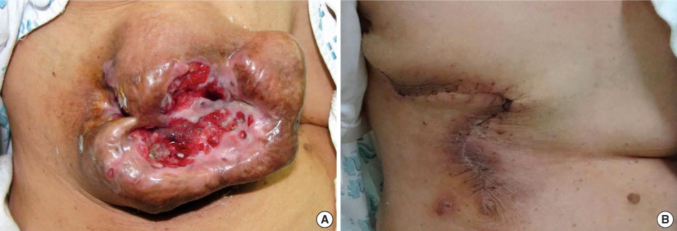
Figure 1 The preoperative (A) and postoperative (B) appearance of the right breast. (A) The necrotized area of the central portion was broad, and it included the nipple and areola. (B) The operative wound was clean without complications after complete excision of the mass and primary repair.
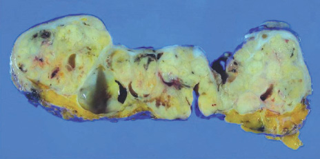
Figure 2 Cut surface of the tumor. It showed multinodularity with fibrous septae and skin invasion that caused ulceration.
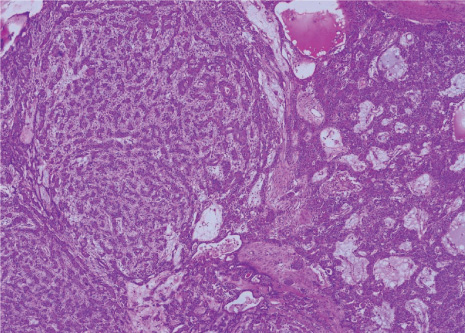
Figure 3 Microscopic finding of the tumor. The tumor showed vaguely nodular growth of proliferating epithelial cells invested by myoepithelial components forming tubules or trabeculae (H&E stain, ×40).
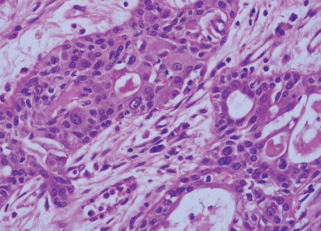
Figure 4 Microscopic finding of the tumor. Cytologic atypia with increased mitotic activity was evident in multiple foci of the glands (H&E stain, ×400).
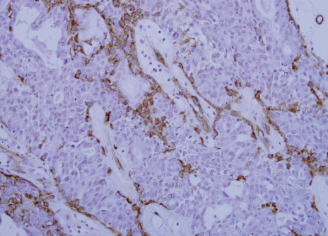
Figure 5 Immunohistochemical staining with alpha-smooth muscle actin. The myoepithelial cells stained for smooth muscle actin (×200).
Reference
-
1. Rosen PP. Adenomyoepithelioma of the breast. Hum Pathol. 1987. 18:1232–1237.
Article2. Yahara T, Yamaguchi R, Yokoyama G, Yamaguchi M, Nakagawa S, Toh U, et al. Adenomyoepithelioma of the breast diagnosed by a mammotome biopsy: report of a case. Surg Today. 2008. 38:144–146.
Article3. Hikino H, Kodama K, Yasui K, Ozaki N, Nagaoka S, Miura H. Intracystic adenomyoepithelioma of the breast: case report and review. Breast Cancer. 2007. 14:429–433.
Article4. Ahmed AA, Heller DS. Malignant adenomyoepithelioma of the breast with malignant proliferation of epithelial and myoepithelial elements: a case report and review of the literature. Arch Pathol Lab Med. 2000. 124:632–636.
Article5. Nadelman CM, Leslie KO, Fishbein MC. "Benign," metastasizing adenomyoepithelioma of the breast: a report of 2 cases. Arch Pathol Lab Med. 2006. 130:1349–1353.
Article6. Hamperl H. The myothelia (myoepithelial cells). Normal state; regressive changes; hyperplasia; tumors. Curr Top Pathol. 1970. 53:161–220.7. Tavassoli FA. Myoepithelial lesions of the breast: myoepitheliosis, adenomyoepithelioma and myoepithelial carcinoma. Am J Surg Pathol. 1991. 15:554–568.8. Iyengar P, Ali SZ, Brogi E. Fine-needle aspiration cytology of mammary adenomyoepithelioma: a study of 12 patients. Cancer. 2006. 108:250–256.
Article9. Clarke LE, Seykora JT. Primary cutaneous adenomyoepithelioma. J Cutan Pathol. 1997. 34:654–657.
Article10. Chang A, Bassett L, Bose S. Adenomyoepithelioma of the breast: a cytologic dilemma. Report of a case and review of the literature. Diagn Cytopathol. 2002. 26:191–196.
Article11. Schürch W, Potvin C, Seemayer TA. Malignant myoepithelioma (myoepithelial carcinoma) of the breast: an ultrastructural and immunocytochemical study. Ultrastruct Pathol. 1985. 8:1–11.
Article12. Chen PC, Chen CK, Nicastri AD, Wait RB. Myoepithelial carcinoma of the breast with distant metastasis and accompanied by adenomyoepitheliomas. Histopathology. 1994. 24:543–548.
Article13. Park MH. Malignant adenomyoepithelioma of the breast. J Korean Surg Soc. 2007. 73:430–433.14. Tavassoli FA, Devilee P. World Health Organization classification of tumours: pathology and genetics of tumours of the breast and female genital organs. 2003. Lyon: IARC Press.15. Bult P, Verwiel JM, Wobbes T, Kooy-Smits MM, Biert J, Holland R. Malignant adenomyoepithelioma of the breast with metastasis in the thyroid gland 12 years after excision of the primary tumor: case report and review of the literature. Virchows Arch. 2000. 436:158–166.
Article
- Full Text Links
-
- Actions
-
Cited
- CITED
-
- Close
- Share
-
- Similar articles
-
- Report of Benign and Malignant Adenomyoepithelioma of the Breast
- Malignant Adenomyoepithelioma of the Breast
- Adenomyoepithelioma of the Breast: Which Mimicking Malignancy on Ultrasound Elastography
- The Breast Adenomyoepithelioma
- Mammographic & Sonographic findings of Adenomyoepithelioma of the Breast: Case Report
