Bone thickness of the infrazygomatic crest area in skeletal Class III growing patients: A computed tomographic study
- Affiliations
-
- 1Department of Orthodontics, College of Dentistry and Research Institute of Oral Science, Gangneung-Wonju National University, Gangneung, Korea. korth@gwnu.ac.kr
- 2Department of Oral and Maxillofacial Radiology, College of Dentistry and Research Institute of Oral Science, Gangneung-Wonju National University, Gangneung, Korea.
- KMID: 2229608
- DOI: http://doi.org/10.5624/isd.2013.43.4.261
Abstract
- PURPOSE
This study was performed to investigate the bone thickness of the infrazygomatic crest area by computed tomography (CT) for placement of a miniplate as skeletal anchorage for maxillary protraction in skeletal Class III children.
MATERIALS AND METHODS
CT images of skeletal Class III children (7 boys, 9 girls, mean age: 11.4 years) were taken parallel to the Frankfurt horizontal plane. The bone thickness of the infrazygomatic crest area was measured at 35 locations on the right and left sides, perpendicular to the bone surface.
RESULTS
The bone was thickest (5.0 mm) in the upper zygomatic bone and thinnest (1.1 mm) in the anterior wall of the maxillary sinus. Generally, there was a tendency for the bone to be thicker at the superior and lateral area of the zygomatic process of the maxilla. There was no clinically significant difference in bone thickness between the right and left sides; however, it was thicker in male than in female subjects.
CONCLUSION
In the infrazygomatic crest area, the superior and lateral area of the zygomatic process of the maxilla had the most appropriate thickness for placement of a miniplate in growing skeletal Class III children with a retruded maxilla.
Figure
-
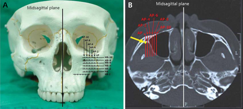
Fig. 1 Construction of the horizontal base plane (HB) and antero-posterior line. A. HB is perpendicular to the midsagittal plane and passes through the most inferior border of the zygomatic process of the maxilla or zygomatic bone. B. The antero-posterior line (AP) is drawn on each horizontal plane parallel to the midsagittal plane and passes through the most anterior point of the infratemporal fossa (arrow).

Fig. 2 Mean of the bilateral bone thickness (mm) map of the infrazygomatic crest area of the zygomatic process of the maxilla. X indicates the bone thickness at each location.

Fig. 3 Male and female bone thickness (mm) maps. X indicates the bone thickness at each point.
Cited by 1 articles
-
Zygomatic miniplates for skeletal anchorage in orthopedic correction of Class III malocclusion: A controlled clinical trial
Erdal Bozkaya, Alime Sema Yüksel, Süleyman Bozkaya
Korean J Orthod. 2017;47(2):118-129. doi: 10.4041/kjod.2017.47.2.118.
Reference
-
1. De Pauw GA, Dermaut L, De Bruyn H, Johansson C. Stability of implants as anchorage for orthopedic traction. Angle Orthod. 1999; 69:401–407.2. Irie M, Nakamura S. Orthopedic approach to severe skeletal Class III malocclusion. Am J Orthod. 1975; 67:377–392.
Article3. Kambara T. Dentofacial changes produced by extraoral forward force in the Macaca irus. Am J Orthod. 1977; 71:249–277.
Article4. Revelo B, Fishman LS. Maturational evaluation of ossification of the midpalatal suture. Am J Orthod Dentofacial Orthop. 1994; 105:288–292.
Article5. Smalley W, Shapiro PA, Hohl TH, Kokich VG, Brånemark PI. Osseointegrated titanium implants for maxillofacial protraction in monkeys. Am J Orthod Dentofacial Orthop. 1988; 94:285–295.
Article6. Roberts WE, Helm FR, Marshall KJ, Gongloff RK. Rigid endosseous implants for orthodontic and orthopedic anchorage. Angle Orthod. 1989; 59:247–256.7. Singer SL, Henry PJ, Rosenberg I. Osseointegrated implants as an adjunct to facemask therapy: a case report. Angle Orthod. 2000; 70:253–262.8. Hong H, Ngan P, Han G, Qi LG, Wei SH. Use of onplants as stable anchorage for facemask treatment: a case report. Angle Orthod. 2005; 75:453–460.9. Cha BK, Lee NK, Choi DS. Maxillary protraction treatment of skeletal Class III children using miniplate anchorage. Korean J Orthod. 2007; 37:73–84.10. Cha BK, Choi DS, Ngan P, Jost-Brinkmann PG, Kim SM, Jang IS. Maxillary protraction with miniplates providing skeletal anchorage in a growing Class III patient. Am J Orthod Dentofacial Orthop. 2011; 139:99–112.
Article11. Cha BK, Ngan PW. Skeletal anchorage for orthopedic correction of growing Class III patients. Semin Orthod. 2011; 17:124–137.
Article12. Sherwood KH, Burch J, Thompson W. Intrusion of supererupted molars with titanium miniplate anchorage. Angle Orthod. 2003; 73:597–601.13. Bengi AO, Karacay S, Akin E, Olmez H, Okçu KM, Mermut S. Use of zygomatic anchors during rapid canine distalization: a preliminary case report. Angle Orthod. 2006; 76:137–147.14. De Clerck H, Geerinckx V, Siciliano S. The zygoma anchorage system. J Clin Orthod. 2002; 36:455–459.15. Seebeck J, Goldhahn J, Städele H, Messmer P, Morlock MM, Schneider E. Effect of cortical thickness and cancellous bone density on the holding strength of internal fixator screws. J Orthop Res. 2004; 22:1237–1242.
Article16. Molly L. Bone density and primary stability in implant therapy. Clin Oral Implants Res. 2006; 17:Suppl 2. 124–135.
Article17. Park CH, Kim KD, Park CS. Measurement of maxillary sinus volume using computed tomography. Korean J Oral Maxillofac Radiol. 2000; 30:63–70.18. Jun BC, Song SW, Park CS, Lee DH, Cho KJ, Cho JH. The analysis of maxillary sinus aeration according to aging process; volume assessment by 3-dimensional reconstruction by high-resolutional CT scanning. Otolaryngol Head Neck Surg. 2005; 132:429–434.
Article19. Lee CH, Rhee CS, Oh SJ, Jun YH, Min YG, Kim IO. Development of the paranasal sinuses in children: a MRI study. Korean J Otolaryngol-Head Neck Surg. 2000; 43:507–513.20. Choi DS, Cha BK, Jang I, Kang KH, Kim SC. Three-dimensional finite element analysis of occlusal stress distribution in the human skull with premolar extraction. Angle Orthod. 2013; 83:204–211.
Article21. Jin GC, Kim KD, Roh BD, Lee CY, Lee SJ. Buccal bone plate thickness of the Asian people. J Endod. 2005; 31:430–434.
Article
- Full Text Links
-
- Actions
-
Cited
- CITED
-
- Close
- Share
-
- Similar articles
-
- Assessment of lower incisor alveolar bone width using cone-beam computed tomography images in skeletal Class III adults of different vertical patterns
- Comparison of mandibular anterior alveolar bone thickness in different facial skeletal types
- A study on relation of position of hyoidbone and upper airway dimensional change according to chin movement in persons with skeletal class III facial pattern after orthognathic surgery
- Location and shape of the mandibular lingula: Comparison of skeletal class I and class III patients using panoramic radiography and cone-beam computed tomography
- A study of the calcification of the second and the third molars in skeletal Class II and III malocclusions
