Comparison of mandibular anterior alveolar bone thickness in different facial skeletal types
- Affiliations
-
- 1Department of Orthodontics, College of Dentisrty, Yonsei University, Korea. hwang@yuhs.ac.
- KMID: 2273222
- DOI: http://doi.org/10.4041/kjod.2010.40.5.314
Abstract
OBJECTIVE
The purpose of this study was to determine differences of mandibular anterior alveolar bone thickness and symphysial cross sectional area in 9 different horizontal and vertical facial types.
METHODS
By using the initial cephalometric radiographs of 270 adult patients (male 135, female 135), the authors measured the buccolingual thickness of anterior alveolar bone on the basis of the root axis and symphysial cross sectional distance.
RESULTS
The high angle group showed significantly thinner buccolingual alveolar bone width except for the CEJ area and lingual alveolar bone width (p < 0.05). The low angle group and Class I, II average group showed similar or significantly thicker alveolar bone width than the Class I average group (p < 0.05). The Class III average group showed significantly thinner buccolingual and lingual alveolar bone width than Class I and II average groups (p < 0.05). The Class III high angle group showed minimal alveolar bone width in all facial skeletal types. No significant difference was found in the symphysial cross sectional area of the different vertical facial skeletal types (p > 0.05).
CONCLUSIONS
The results of this study found that Class III high angle patients have thinner mandibular anterior alveolar bone thickness; therefore, more attention will be needed to determine the incisor position during orthodontic treatment for this group of patients.
Keyword
Figure
-
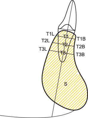
Fig. 1 Measurement of mandibular alveolar bone width and dimension. T1T, Buccolingual width (mm) of mandibular anterior alveolar bone at 2 mm under CEJ; T2T, buccolingual width (mm) of mandibular alveolar bone at middle of root; T3T, buccolingual width (mm) of mandibular alveolar bone at 2 mm over root apex; T1L, lingual alveolar bone thickness (mm) of mandible at 2 mm under CEJ; T2L, lingual alveolar bone thickness (mm) of mandible at middle of root; T3L, lingual alveolar bone thickness (mm) of mandible at 2 mm over root apex; T1B, buccal alveolar bone thickness (mm) of mandible at 2 mm under CEJ; T2B, buccal alveolar bone thickness (mm) of mandible at middle of root; T3B, buccal alveolar bone thickness (mm) of mandible at 2 mm over root apex; S, symphysial cross sectional area (mm2) of including root area.
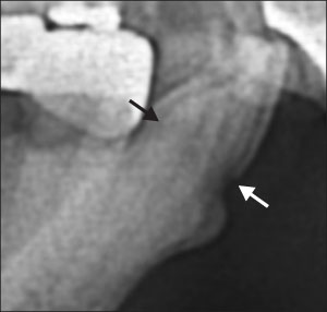
Fig. 2 Buccal and lingual cemento-enamel junction (CEJ) on cephalometric X-ray film.
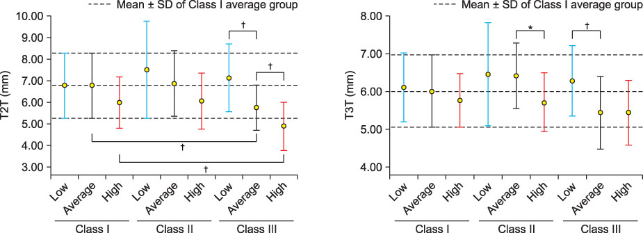
Fig. 3 Error bar graph (Mean ± SD) of buccolingual alveolar bone width at midroot (T2T) and root apex (T3T). *Significant at the significance level of 0.05; †significant at the significance level of 0.01.

Fig. 4 Error bar graph (Mean ± SD) of lingual alveolar bone width at CEJ (T1L), midroot (T2L), and root apex (T3L). *Significant at the significance level of 0.01.
Cited by 3 articles
-
Correction of dental Class III with posterior open bite by simple biomechanics using an anterior C-tube miniplate
Hyo-Won Ahn, Kyu-Rhim Chung, Suk-Man Kang, Lu Lin, Gerald Nelson, Seong-Hun Kim
Korean J Orthod. 2012;42(5):270-278. doi: 10.4041/kjod.2012.42.5.270.Assessment of lower incisor alveolar bone width using cone-beam computed tomography images in skeletal Class III adults of different vertical patterns
Sanghee Lee, Soonshin Hwang, Woowon Jang, Yoon Jeong Choi, Chooryung J Chung, Kyung-Ho Kim
Korean J Orthod. 2018;48(6):349-356. doi: 10.4041/kjod.2018.48.6.349.Alveolar bone thickness and fenestration of incisors in untreated Korean patients with skeletal class III malocclusion: A retrospective 3-dimensional cone-beam computed tomography study
Song Hee Oh, Kyung-Yen Nahm, Seong-Hun Kim, Gerald Nelson
Imaging Sci Dent. 2020;50(1):9-14. doi: 10.5624/isd.2020.50.1.9.
Reference
-
1. Edwards JG. A study of the anterior portion of the palate as it relates to orthodontic therapy. Am J Orthod. 1976. 69:249–273.
Article2. Ten Hoeve A, Mulie RM. The effect of antero-postero incisor repositioning on the palatal cortex as studied with laminagraphy. J Clin Orthod. 1976. 10:804–822.3. Handelman CS. The anterior alveolar: its importance in limiting orthodontic treatment and its influence on the occurrence of iatrogenic sequelae. Angle Orthod. 1996. 66:95–109.4. Graber TM, Vanarsdall RL. Orthodontics: current principles and techniques. 1994. 2nd ed. St Louis: Mosby.5. Wehrbein H, Bauer W, Diedrich P. Mandibular incisors, alveolar bone, and symphysis after orthodontic treatment. A retrospective study. Am J Orthod Dentofacial Orthop. 1996. 110:239–246.
Article6. Vardimon AD, Oren E, Ben-Bassat Y. Cortical bone remodeling/tooth movement ratio during maxillary incisor retraction with tip versus torque movements. Am J Orthod Dentofacial Orthop. 1998. 114:520–529.
Article7. Choe HY, Park W, Jeon JK, Kim YH, Shon BW. Differences in mandibular anterior alveolar bone thickness according to age in a normal skeletal group. Korean J Orthod. 2007. 37:220–230.8. Sarikaya S, Haydar B, Ciğer S, Ariyürek M. Changes in alveolar bone thickness due to retraction of anterior teeth. Am J Orthod Dentofacial Orthop. 2002. 122:15–26.
Article9. Wainwright WM. Faciolingual tooth movement: its influence on the root and cortical plate. Am J Orthod. 1973. 64:278–302.
Article10. Remmelink HJ, van der Molen AL. Effect of anteroposterior incisor repositioning on the root and cortical plate: a follow-up study. J Clin Orthod. 1984. 18:42–49.11. Wehrbein H, Fuhrmann RA, Diedrich PR. Periodontal conditions after facial root tipping and palatal root torque of incisors. Am J Orthod Dentofacial Orthop. 1994. 106:455–462.
Article12. Gündüz E, Rodríguez-Torres C, Gahleitner A, Heissenberger G, Bantleon HP. Bone regeneration by bodily tooth movement: dental computed tomography examination of a patient. Am J Orthod Dentofacial Orthop. 2004. 125:100–106.
Article13. Larato DC. Alveolar plate fenestrations and dehiscence of the human skull. Oral Surg Oral Med Oral Pathol. 1970. 29:816–819.14. Nauert K, Berg R. Evaluation of labio-lingual bony support of lower incisors in orthodontically untreated adults with the help of computed tomography. J Orofac Orthop. 1999. 60:321–334.
Article15. Ha YR. Comparison of anterior alveolar bone thickness and resorption patterns on adults and adolescents due to retraction of anterior teeth in mandibule. 2006. Seoul: The Graduate School of Yonsei University.16. Li JL, Li XB, Li JY, Qiao J, Peng MH, Qian X. Study of mandibular anterior alveolar bone thickness in subjects with different facial skeletal types. Hua Xi Kou Qiang Yi Xue Za Zhi. 2008. 26:399–401.17. Chung CJ, Jung S, Baik HS. Morphological characteristics of the symphyseal region in adult skeletal Class III crossbite and openbite malocclusions. Angle Orthod. 2008. 78:38–43.
Article18. Reitan K. The tissue reaction as related to the functional factor. Dent Rec (London). 1951. 71:173–183.
Article19. Reitan K. Initial tissue behavior during apical root resorption. Angle Orthod. 1974. 44:68–82.20. Douglass C, Gillings D, Sollecito W, Gammon M. National trends in the prevalence and severity of the periodontal diseases. J Am Dent Assoc. 1983. 107:403–412.
Article21. Van der Velden U. Effect of age on the periodontium. J Clin Periodontol. 1984. 11:281–294.
Article22. Sched O, Waerhaug J, Lovdal A, Arno A. Alveolar bone loss as related to oral hygiene and age. J Periodontol. 1959. 30:7–16.
Article23. Harris EF, Dyer GS, Vaden JL. Age effects of orthodontic treatment: skeletodental assessments from the Johnston analysis. Am J Orthod Dentofacial Orthop. 1991. 100:531–536.
Article24. Wehrbein H, Fuhrmann RA, Diedrich PR. Human histologic tissue response after long-term orthodontic tooth movement. Am J Orthod Dentofacial Orthop. 1995. 107:360–371.
Article25. Fuhrmann R. Three-dimensional interpretation of labiolingual bone width of the lower incisors. Part II. J Orofac Orthop. 1996. 57:168–185.
- Full Text Links
-
- Actions
-
Cited
- CITED
-
- Close
- Share
-
- Similar articles
-
- Differences in mandibular anterior alveolar bone thickness according to age in a normal skeletal group
- Angulation between Long Axis of Anterior Teeth and Alveolar Process, and Thickness of Alveolar Bone
- Assessment of lower incisor alveolar bone width using cone-beam computed tomography images in skeletal Class III adults of different vertical patterns
- Comparison of anterior maxillary and mandibular alveolar parameters in African American and Caucasian women: A retrospective pilot study
- A roentgenocephalometric study of the bony structure and its profile
