Successful Pacemaker Revision Through Sustained Right Superior Vena Cava in a Patient With Situs Inversus Totalis
- Affiliations
-
- 1Department of Internal Medicine, Seoul National University College of Medicine, Seoul, Korea. seil@snu.ac.kr
- KMID: 2225855
- DOI: http://doi.org/10.4070/kcj.2008.38.2.128
Abstract
- In patients with situs inversus totalis, the superior vena cava is normally positioned on the left side and drains into a left-sided right atrium (RA). If right-side superior vena cava (RSVC) is also present, it should be thought of as a combined congenital anomaly. Here, we report a case of successful pacemaker lead insertion through the RSVC in a patient with situs inversus totalis. The left-side superior vena cava (LSVC) had been already used as a route for the first pacemaker lead insertion 15 years earlier. During the pacemaker lead revision, we found that the LSVC was obliterated, and used the RSVC as a route for a new pacemaker lead insertion.
Figure
-

Fig. 1 Initial chest X-ray on admission. The chest X-ray showed situs inversus totalis.
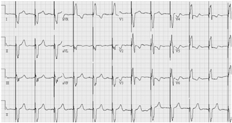
Fig. 2 Initial ECG on admission. Tall pacing spikes resulted from ventricular pacing with high output. ECG: electrocardiography.

Fig. 3 Pacemaker revision through RSVC. In Figure A, the previously inserted pacemaker lead is shown in the LSVC (arrows). However, the LSVC was not visualized below the level of the most inferior arrow in this figure, and we could not pass a guide wire in order to insert a new pacemaker lead. However, the RSVC drained into the right atrium via the coronary sinus, and we were able to insert a guide wire into the left-sided right ventricle (B). Arrowheads indicate the venous drainage route to the RSVC. LSVC: left superior vena cava, RSVC: right superior vena cava.

Fig. 4 Chest X-ray after the procedure. The chest X-ray after successful pacemaker revision showed that the new pacemaker lead (arrowheads) was positioned in the right ventricular apex through the RSVC. RSVC: right superior vena cava.

Fig. 5 ECG during the 1-month follow-up after pacemaker revision. This ECG shows a well-functioning pacemaker. ECG: electrocardiography.
Reference
-
1. Biffi M, Boriani G, Frabetti L, Bronzetti G, Branzi A. Left superior vena cava persistence in patients undergoing pacemaker or cardioverter-defibrillator implantation: a 10-year experience. Chest. 2001. 120:139–144.2. Murayama H, Maeda M, Sakurai H, Watanabe T. Absent left superior vena cava with persistent right superior vena cava in visceroatrial situs inversus. Pediatr Cardiol. 2006. 27:293–296.3. Son JW, Lee CS, Han SW, Lee SW, Kim SK, Kwon YJ. A case of persistent left SVC associated with tricuspid regurgitation. Korean Circ J. 1993. 23:609–613.4. Campbell M, Deuchar DC. The left-sided superior vena cava. Br Heart J. 1954. 16:423–439.5. Morgan DR, Hanratty CG, Dixon LJ, Trimble M, O'Keeffe DB. Anomalies of cardiac venous drainage associated with abnormalities of cardiac conduction system. Europace. 2002. 4:281–287.6. Nsah EN, Moore GW, Hutchins GM. Pathogenesis of persistent left superior vena cava with a coronay sinus connection. Pediatr Pathol. 1991. 11:261–269.7. Rigatelli G. Congenitally persistent left superior vena cava: a possible unpleasant problem during invasive procedures. J Cardiovasc Med (Hagerstown). 2007. 8:483–487.8. Raghib G, Ruttenberg HD, Anderson RC, Amplatz K, Adams P Jr, Edwards JE. Termination of left superior vena cava in left atrium, atrial septal defect, and absence of coronary sinus: a developmental complex. Circulation. 1965. 31:906–918.9. Jeong SY, Sin PJ, Cho SY, et al. A case of situs inversus (I.D.D.) with corrected TGA. Korean Circ J. 1993. 23:296–301.10. Gonzalez-Juanatey C, Testa A, Vidan J, et al. Persistent left superior vena cava draining into thecoronary sinus: report of 10 cases and literature review. Clin Cardiol. 2004. 27:515–518.11. Zaglavara T, Hamilton JR, Kenny A. A combination of persistent left superiorvena cava and a large secundumatrial septal defect in a 34 year old woman. Heart. 2001. 85:406.12. Gheissari A, Malm JR, Bowman FO Jr, Bierman FZ. Cor triatriatumsinistrum: one institution's 28-year experience. Pediatr Cardiol. 1992. 13:85–88.13. Krukal JC. Transvenous pacemaker failure due to anomalous venous return to the heart. Chest. 1971. 59:458–461.14. Garcia L, Levine R, Kosowsky W, Lyon AF. Persistent left superior vena cava complicating pacemaker insertion. Chest. 1972. 61:396–397.15. Rubenfire M, Evangelista J, Wajszczuk WJ, Kantrowitz A. Implication of a persistent left superior vena cava in transvenous pacemaker therapy and cardiac hemodynamic monitoring. Chest. 1974. 65:145–147.
- Full Text Links
-
- Actions
-
Cited
- CITED
-
- Close
- Share
-
- Similar articles
-
- Single Port Laparoscopic Cholecystectomy in a Patient with Situs Inversus Totalis: A Case Report
- Neonatal Duodenal Obstruction Associated with Situs Inversus Totalis: A Case Report
- Laparoscopic Low Anterior Resection in a Rectal Cancer Patient with Situs Inversus Totalis: A Case Report
- Laparoscopic cholecystectomy in a case of situs inversus totalis: a review of technical challenges and adaptations
- Radical Subtotal Gastrectomy in Early Gastric Cancer Patient with Situs Inversus Totalis
