Diagnostic Value of Ultrasound-Based Strain Imaging in Patients With Suspected Coronary Artery Disease
- Affiliations
-
- 1Division of Cardiology, Maryknoll Medical Center, Busan, Korea. Kyoungim74@dreamwiz.com
- KMID: 2225759
- DOI: http://doi.org/10.4070/kcj.2008.38.8.398
Abstract
-
BACKGROUND AND OBJECTIVES: Strain imaging has already been shown to quantify regional myocardial function in both acute ischemic myocardium and infarcted myocardium. We proposed that strain imaging could differentiate deformation of normal and ischemic myocardium that are without regional wall motion abnormality, as assessed by conventional echocardiography. The aim of this study is to determine the diagnostic value of strain imaging for the detection and localization of coronary lesions in patients with chest pain, but they are without apparent wall motion abnormalities.
SUBJECTS AND METHODS
Strain imaging for advanced wall motion analysis was performed in 179 patients with suspicious stable angina (SA) and in 94 patients with suspicious acute coronary syndrome (ACS) prior to coronary angiography. All the patients had normal conventional wall motion scoring based on the standards of the American Society of Echocardiography. Longitudinal strain was measured in 3 apical views, and assessments of the strain value for individual segments with using an 18-segment division of the left ventricle were performed to determine the average strain value. Marked heterogeneity of strain was considered abnormal, and significant coronary artery disease was considered present if stenosis above 70% was noted on the quantitative angiography.
RESULTS
Eighty (78%) of the 103 patients with SA and 18 (56%) of the 32 patients with ACS and who showed constant systolic strain throughout the left ventricular wall had normal or minimal coronary lesions. Fifty-one (67%) of the 76 patients with SA and 53 (85%) of the 62 patients with ACS and marked heterogeneity of strain had angiographically significant coronary stenosis. The receiver-operating characteristic (ROC) analysis of the peak systolic strain yielded that the ROC-area of peak systolic strain for the left anterior descending artery territory was 0.79 (95% CI 0.72-0.84), this was 0.87 (95% CI 0.79-0.91) for the left circumflex artery territory and 0.89 (95% CI 0.79-0.93) for the right coronary artery territory.
CONCLUSION
Ultrasound-based strain imaging demonstrates a strong correlation with coronary angiography and it has potential as a noninvasive diagnostic tool for detecting coronary artery stenosis in patients with chest pain, but who are without apparent wall motion abnormalities on conventional echocardiography.
Keyword
MeSH Terms
Figure
-
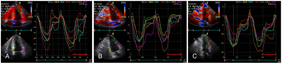
Fig. 1 Strain echocardiography in a patient with normal angiography shows a relatively homogeneous pattern of peak systolic strains throughout the LV in the apical 4-chamber view (A), the apical 2-chamber view (B) and the apical 3-chamber view (C). LV: left ventricle.
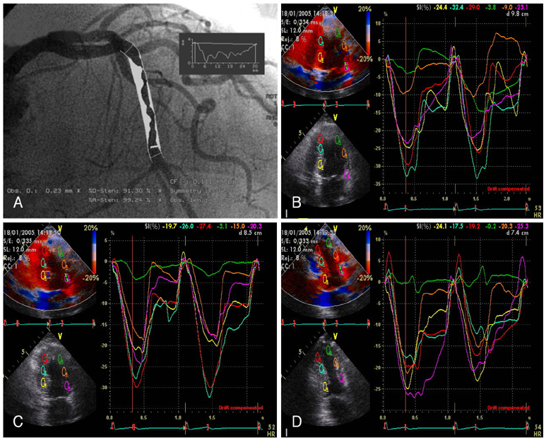
Fig. 2 Strain echocardiography in a patient with significant left anterior descending coronary artery stenosis (A) shows a marked heterogeneous pattern of peak systolic strains throughout the LV in the apical 4-chamber view (B), the apical 2-chamber view (C) and the apical 3-chamber view (D). LV: left ventricle.

Fig. 3 Strain echocardiography in a patient with significant left anterior descending coronary artery stenosis (A) and right coronary artery stenosis (B) shows a marked heterogeneous pattern of peak systolic strains throughout the LV in the apical 3-chamber view (C) and the apical 2-chamber view (D). LV: left ventricle.
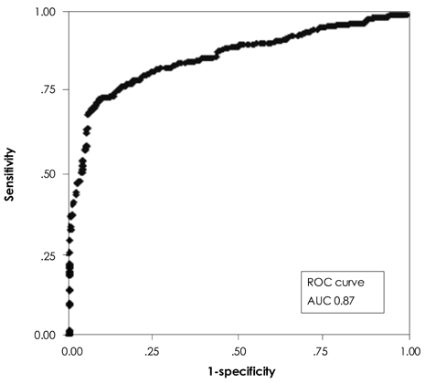
Fig. 4 Receiver-operating characteristic curves for peak systolic strain. AUC: area under the curve, ROC: receiver operating characteristics.
Reference
-
1. Hoffman R, Lethen H, Marwick T, et al. Analysis of interinstitutional observer agreement in interpretation of dobutamine stress echocardiograms. J Am Coll Cardiol. 1996. 27:330–336.2. Kvitting JP, Wigstrom L, Strotmann JM, Sutherland GR. How accurate is visual assessment of synchronicity in myocardial motion?: an in vitro study with computer-simulated regional delay in myocardial motion: clinical implications forrest and stress echocardiography studies. J Am Soc Echocardiogr. 1999. 12:698–705.3. Ehring T, Heusch G. Left ventricular asynchrony: an indicator of regional myocardial dysfunction. Am Heart J. 1990. 120:1047–1057.4. Osakada G, Hess OM, Gallagher KP, Kemper WS, Ross J. End-systolic dimension-wall thickness relations during myocardial ischemia in conscious dogs: a new approach for defining regional function. Am J Cardiol. 1983. 51:1750–1758.5. Wiegner AW, Allen GJ, Bing OH. Weak and strong myocardium in series: implications for segmental dysfunction. Am J Physiol. 1978. 235:H776–H783.6. Leone BJ, Norris RM, Safwat A, Foex P, Ryder WA. Effects of progressive myocardial ischaemia on systolic function, diastolic dysfunction, and load dependent relaxation. Cardiovasc Res. 1992. 26:422–429.7. Derumeaux G, Ovize M, Loufoua J, et al. Doppler tissue imaging quantitates regional wall motion during myocardial ischemia and reperfusion. Circulation. 1998. 97:1970–1977.8. Derumeaux G, Ovize M, Loufoua J, Pontier G, Andre-Fouet X, Cribier A. Assessment of nonuniformity of transmural myocardial velocities by color-coded tissue Doppler imaging: characterization of normal, ischemic, and stunned myocardium. Circulation. 2000. 101:1390–1395.9. Armstrong G, Pasquet A, Fukamachi K, Cardon L, Olstad B, Marwick T. Use of peak systolic strain as an index of regional left ventricular function: comparison with tissue Doppler velocity during dobutamine stress and myocardial ischemia. J Am Soc Echocardiogr. 2000. 13:731–737.10. Urheim S, Edvardsen T, Torp H, Angelsen B, Smiseth O. Myocardial strain by Doppler echocardiography: validation of a new method to quantify regional myocardial function. Circulation. 2000. 102:1158–1164.11. Mirsky I, Parmley WW. Assessment of passive elastic stiffness for isolated heart muscle and the intact heart. Circ Res. 1973. 33:233–243.12. Heimdal A, Stoylen A, Torp H, Skaerpe T. Real-time strain rate imaging of the left ventricle by ultrasound. J Am Soc Echocardiogr. 1998. 11:1013–1019.13. Tennant R, Wiggers CJ. The effect of coronary occlusion on myocardial contraction. Am J Physiol. 1935. 112:351–361.14. Visser CA, David GK, Kan G, et al. Two-dimensional echocardiography during percutaneous transluminal coronary angioplasty. Am Heart J. 1986. 111:1035–1041.15. Wohlgelernter D, Cleman M, Highman HA, et al. Regional myocardial dysfunction during coronary angioplasty: evaluation by two-dimensional echocardiography and 12 lead electrocardiography. J Am Coll Cardiol. 1986. 7:1245–1254.16. Voigt JU, Arnold M, Karlsson M, et al. Assessment of regional longitudinal myocardial strain rate derived from Doppler myocardial imaging indexes in normal and infarcted myocardium. J Am Soc Echocardiogr. 2000. 13:588–598.17. Kukulski T, Jamal F, D'Hooge J, Bijnens B, De Scheerder I, Sutherland G. Acute changes in systolic and diastolic events during clinical coronary angioplasty: a comparison of regional velocity, strain rate and strain measurement. J Am Soc Echocardiogr. 2002. 15:1–12.18. Cho KI, Park JH, Park JR, et al. Assessment of left ventricular function in symptomatic patients with myocardial bridge using two-dimensional strain. Korean Circ J. 2006. 36:617–625.19. Stoylen A, Heimdal A, Bjornstard K, et al. Strain rate imaging by ultrasonography in the diagnosis of coronary artery disease. J Am Soc Echocardiogr. 2000. 13:1053–1064.20. Henein MY, O'Sullivan CH, Davies SW, Sigwart U, Gibson DG. Effects of acute coronary occlusion and previous ischemic injury on left ventricular wall motion in humans. Heart. 1997. 77:338–345.21. Cho GY, Park WJ, Han SW, et al. Quantification of regional wall motion abnormality using myocardial strain in acute myocardial infarction. Korean Circ J. 2003. 33:583–589.22. Jamal F, Kukulski T, Strottman JM, et al. Quantitation of the spectrum of changes in regional myocardial function during acute ischemia in closed chest pigs: an ultrasonic strain rate and strain study. J Am Soc Echocardiogr. 2001. 14:874–884.
- Full Text Links
-
- Actions
-
Cited
- CITED
-
- Close
- Share
-
- Similar articles
-
- The Use of Cardiac Magnetic Resonance in Patients with Suspected Coronary Artery Disease: A Clinical Practice Perspective
- Practical Application of Coronary Imaging Devices in Cardiovascular Intervention
- Stress Testing and Imaging Protocols for Myocardial Perfusion Studies
- Carotid ultrasound in patients with coronary artery disease
- Assessment of Myocardial Ischemia Using Stress Perfusion Cardiovascular Magnetic Resonance
