A Case of Conjunctival Inclusion Cyst Managed with Marsupialization
- Affiliations
-
- 1Department of Ophthalmology, Hallym University College of Medicine, Anyang, Korea. minjoung@hallym.or.kr
- KMID: 2218425
- DOI: http://doi.org/10.3341/jkos.2014.55.2.289
Abstract
- PURPOSE
We present a case with conjunctival inclusion cyst at inferior fornix treated by marsupialization.
CASE SUMMARY
A 23-year-old woman visited our clinic complaining of left lower eyelid swelling. Ophthalmologic examination and CT scan showed a cystic mass from inferior conjunctival fornix to anterior orbit with shallow fornix and focal symblepharon. The cyst was effectively removed with marsupializaion. Postoperatively, there was no recurrence of cyst and the fornix was deepened.
CONCLUSIONS
Marsupialization can be a considerable treatment option in conjunctival inclusion cyst, especially when accompanied by shallow fornix and symblepharon.
Figure
-
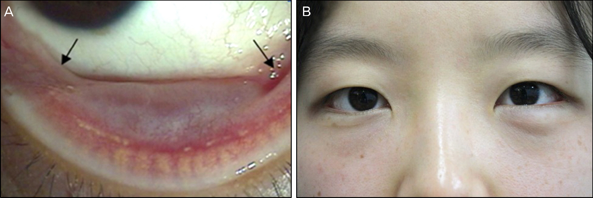
Figure 1. Anterior segment photograph of conjunctival inclusion cyst at initial presentation. (A) About 1.5 × 1 cm sized subcon-junctival cyst is noted at inferior fornix in the left eye. There is symblepharon at medial and lateral end of cyst (black arrows). (B) External photographs showed contour of the mass, which is transcutaneously visible at left lower eyelid.
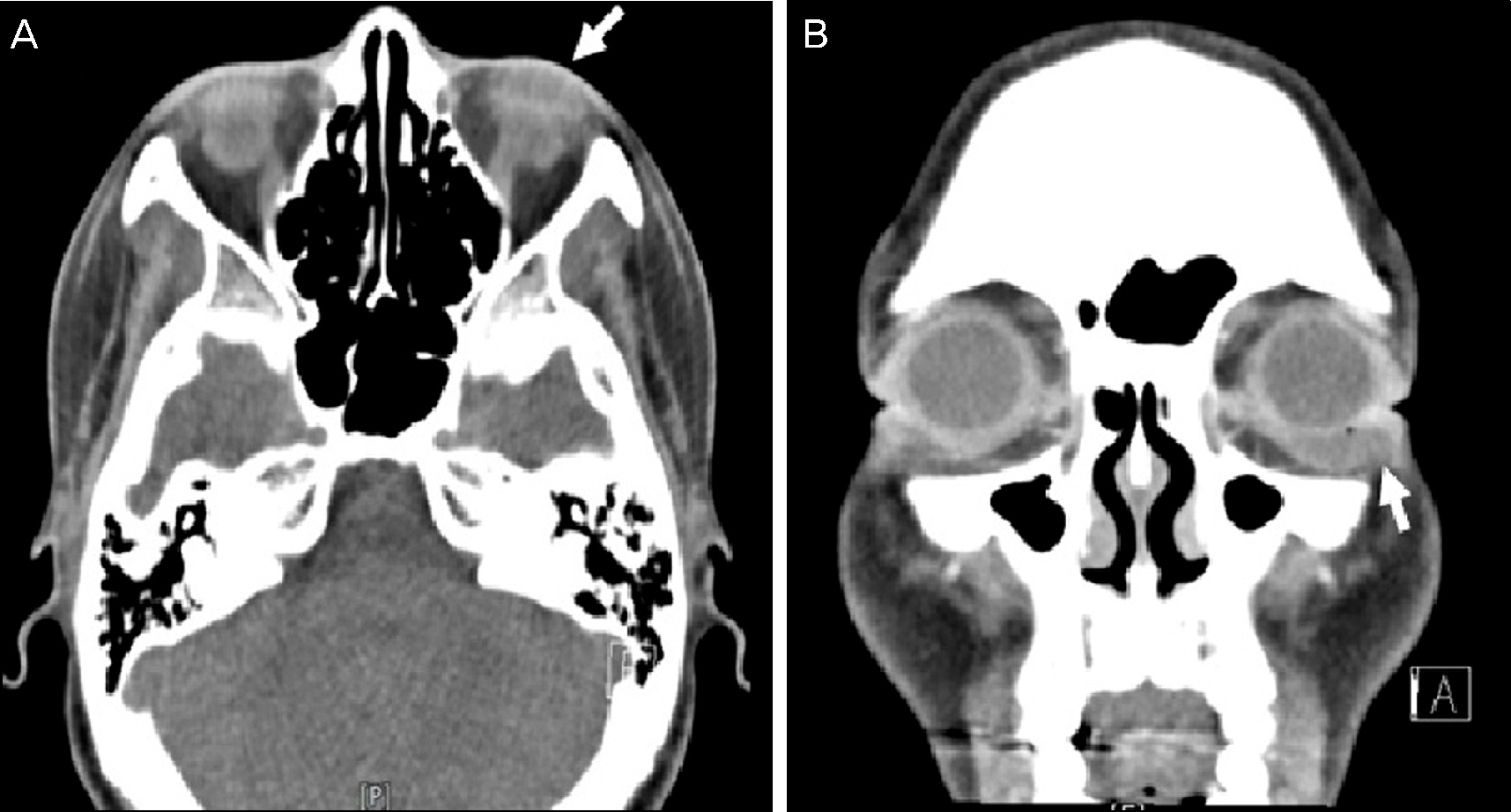
Figure 2. Axial (A) and coronal (B) view enhanced computed tomography images demonstrate 1.5 × 0.8 cm sized ovoid shaped cystic mass (white arrow) in anterior– inferior part of the left orbit.
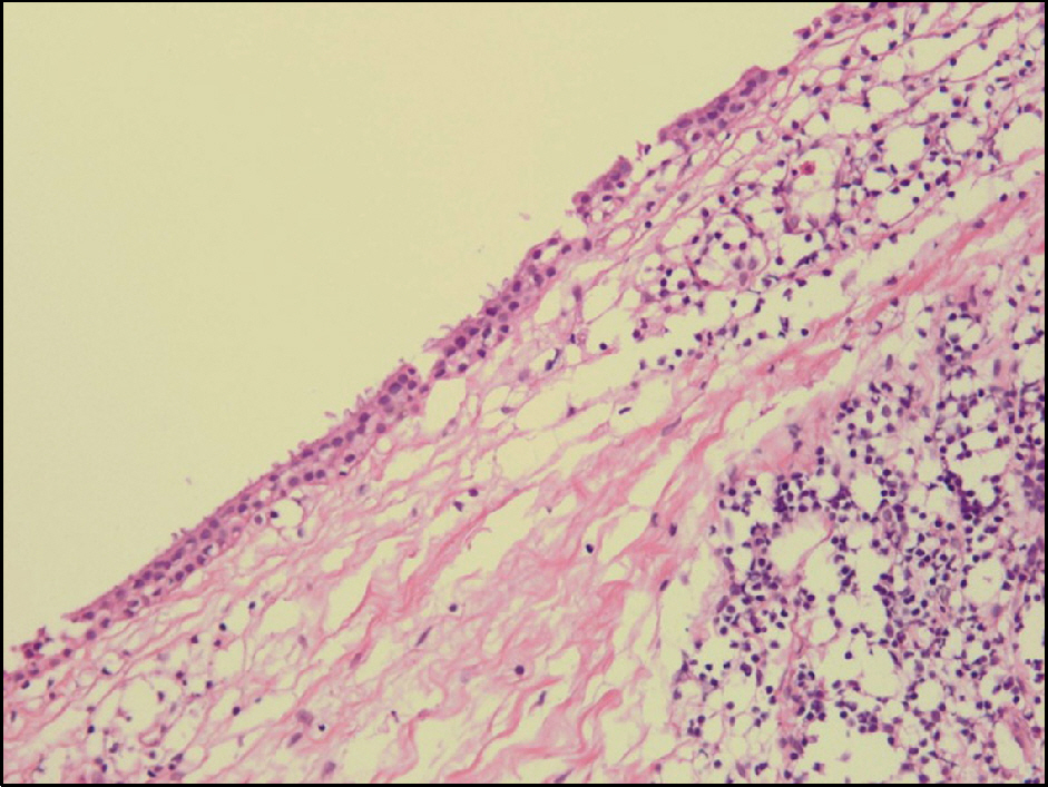
Figure 3. Microscopic appearance of the cyst lined by non-keratinized stratified squamous epithelium with chronic inflammation (H&E, ×100).
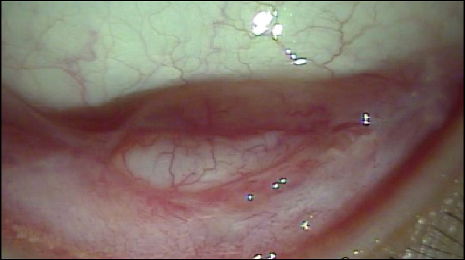
Figure 4. Anterior segment photograph at 1 month after surgery. The posterior wall of the inclusion cyst is covering inferior fornix. There is no evidence of recurrence.
Reference
-
References
1. Sameshima SS, Beyer-Machule CK.Acquired ptosis associated with a conjunctival cyst. Ophthal Plast Reconstr Surg. 1988; 4:159–62.
Article2. Memarzadeh F, Chuck RS, McCulley TJ.Fornix reconstruction with conjunctival inclusion cyst marsupialization in Stevens-Johnson syndrome. Ophthal Plast Reconstr Surg. 2006; 22:475–6.
Article3. Goodglick TA, Mertz P, Wolfley D. . Ciliated respiratory-like epithelium forming cystic conjunctival lesions in a patient with Stevens-Johnson syndrome. Ophthalmic Surg. 1992; 23:557–9.
Article4. Lee J, Kwak AY, Chung WS, Ha BJ.A new simple technique for re-moval of subconjunctival cyst under the slit lamp microscope. J Korean Ophthalmol Soc. 2011; 52:1531–6.
Article5. Kim B, Kang NY.Successful removal of apocrinehydrocytoma us-ing indocyanine green and sodium hyaluronate. J Korean Ophthalmol Soc. 2011; 52:994–8.
Article6. Desai VN, Shields CL, Shields JA.Orbital cyst in a patient with Stevens-Johnson syndrome. Cornea. 1992; 11:592–4.
Article7. Harris GJ.Marsupialization of a lacrimal gland cyst. Ophthalmic Surg. 1983; 14:75–8.
Article8. McCulley TJ, Kersten RC, Yip CC, Kulwin DR.Dacryocystoceles in the aftermath of Stevens-Johnson syndrome. Ophthal Plast Reconstr Surg. 2005; 21:159–61.
Article
- Full Text Links
-
- Actions
-
Cited
- CITED
-
- Close
- Share
-
- Similar articles
-
- Transnasal Marsupialization of Large Infected Radicular Cyst in Immunocompromised Patients: Case Report
- A Case of The Surgically Treated Intraspinal Extradural Meningeal Cyst Demonstrating 'Ball-Valve' Mechanism of Formation
- Retroperitoneal Laparoscopic Marsupialization of Simple Renal Cyst
- A Case of Conjunctival Dermoid Cyst of the Orbit
- A Case of Nasolacrimal Duct Obstruction Caused by a Lacrimal Sac Retention Cyst
