A Case of Conjunctival Dermoid Cyst of the Orbit
- Affiliations
-
- 1Department of Ophthalmology, Catholic University of Daegu College of Medicine, Daegu, Korea. kimkh@cu.ac.kr
- KMID: 2213405
- DOI: http://doi.org/10.3341/jkos.2010.51.4.601
Abstract
- PURPOSE
To report a case of primary conjunctival dermoid of the superonasal orbit.
CASE SUMMARY
A 41-year-old man complained of swelling of the left lower eyelid and left periocular pain for a week. Examination revealed 3 mm of proptosis with superotemporal displacement of the left eye. Orbital CT revealed a 32x27x33-mm well-defined giant cyst with a fat-fluid level in the superonasal aspect of the left orbit. Orbital MRI showed bone remodeling around the cyst, consistent with a dermoid cyst. The cyst was approached via lateral orbitotomy and transcaruncular incision but was ruptured just prior to the end of the dissection and was totally excised using a cryoprobe to freeze the ruptured site. Upon histopathological examination, the cyst was misdiagnosed as a conjunctival cyst because there was no dermal appendage but was rediagnosed as a conjunctival dermoid cyst after the tissue sample was examined more thoroughly. After surgery, the patient presented with diplopia due to esodeviation and was prescribed prismatic lenses.
MeSH Terms
Figure
-
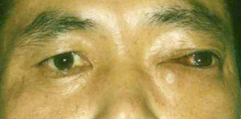
Figure 1. External photograph of a 41-year-old man with progressive proptosis and lateral displacement of the left eye.
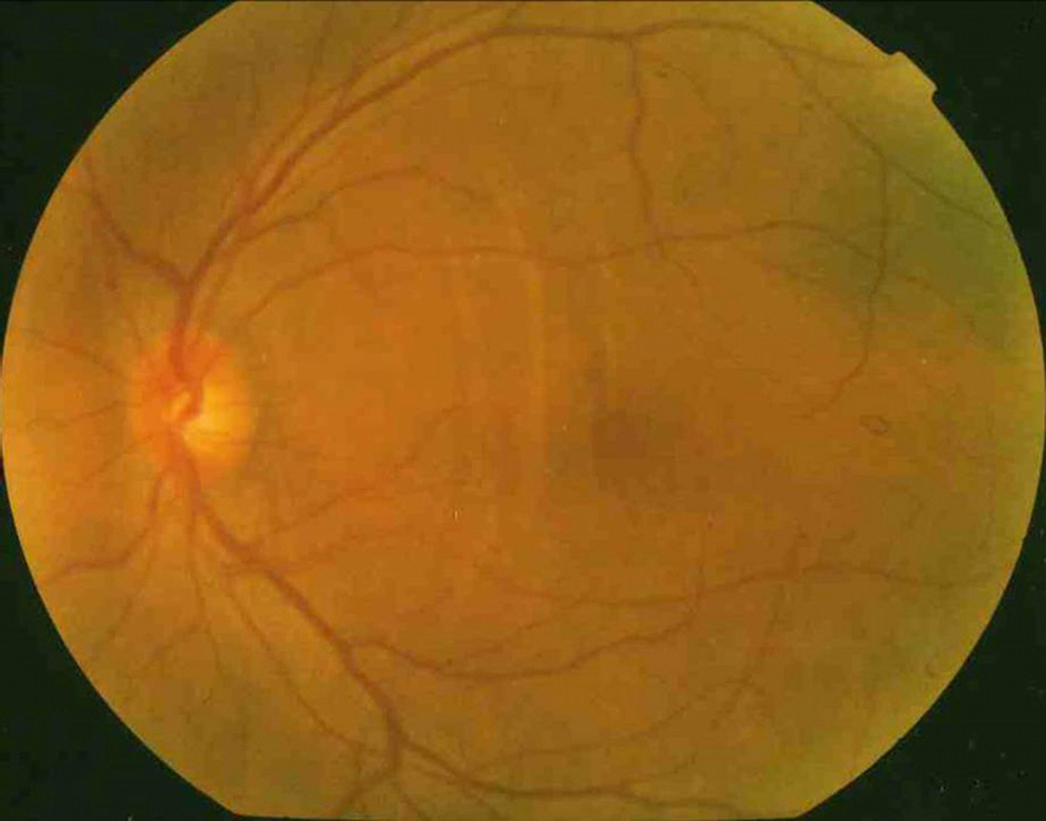
Figure 2. Fundus photograph of the left eye showing choroidal folds at the posterior pole due to globe compression.

Figure 3. Preoperative axial (A) and coronal views (B) of orbital CT scan showing a well-defined unilocular giant cyst (red arrow), measuring 32×27×33 mm, occupying the superonasal area of the left orbit.

Figure 4. Preoperative axial view of orbital MRI reveals a well-defined cyst of the nasal orbit showing high signal intensity on T1-(left) and relatively homogenous enhanced on post-contrast fat suppressed T1-weighted image (right). Globe compression and bone remodeling are seen.
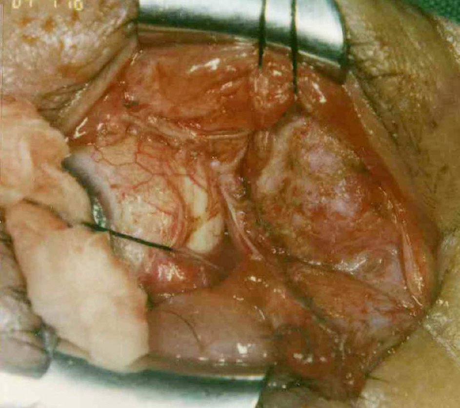
Figure 5. Surgical photograph demonstrating the conjunctival dermoid cyst in the left orbit.
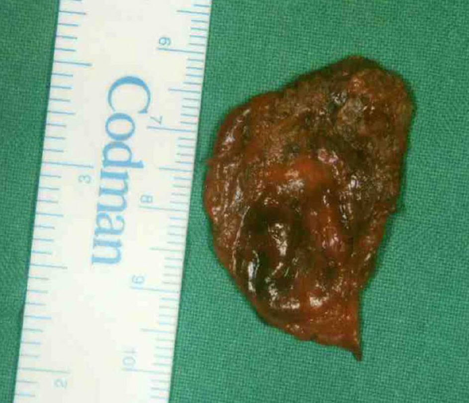
Figure 6. Gross finding of the completely excised conjunctival dermoid cyst which was accidentally perfo-rated and aspirated during operation.
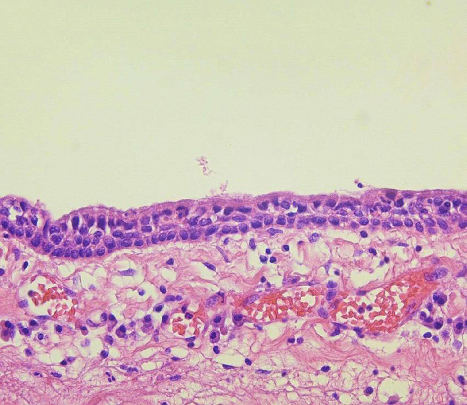
Figure 7. Microscopic examination of cyst wall shows a triple layer of nonkeratinizing stratified epithelium with underlying loose connective tissue. Definite goblet cells and dermal appendages are not seen (hematoxylin and eosin stain, ×400).
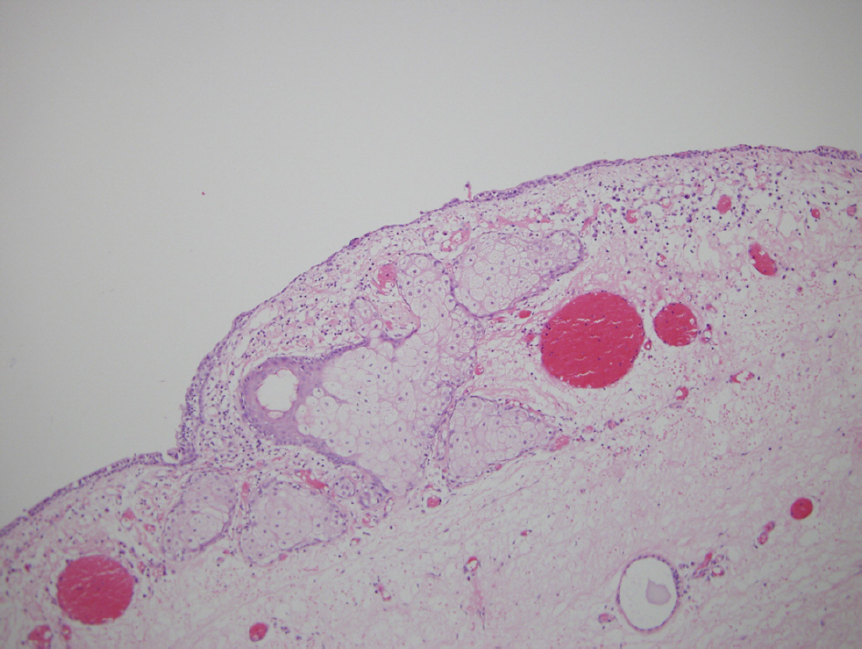
Figure 8. Microscopic examination of cyst wall. The cyst is lined by nonkeratizing epithelium and has dermal appendages (sebaceous glands) (hematoxylin and eosin stain, ×200).
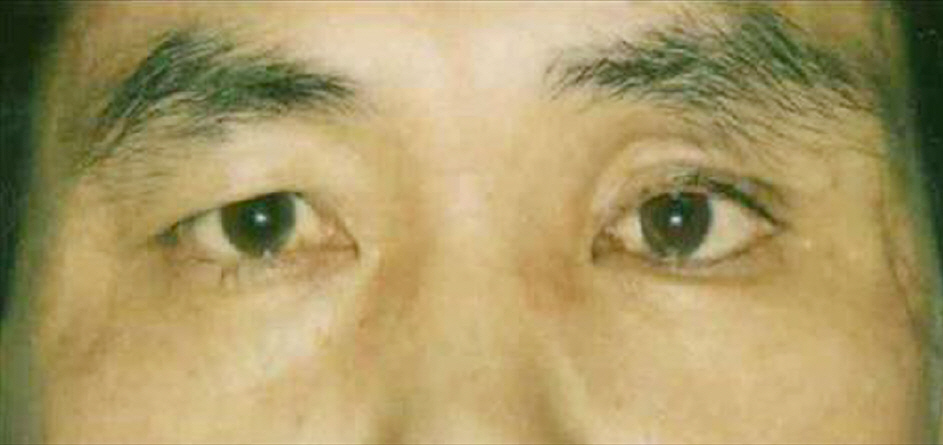
Figure 9. External photograph showing mild esodeviation and enophthalmos in the left eye 2 months after surgery.
Cited by 1 articles
-
Clinical Features of Conjunctival Dermolipoma
Hee Jun Song, Ho Sik Hwang, Yoon Yang Jung, Ji Won Kwon
J Korean Ophthalmol Soc. 2018;59(12):1108-1113. doi: 10.3341/jkos.2018.59.12.1108.
Reference
-
References
1. Shields JA, Augsburger JJ, Donoso LA. Orbital dermoid cyst of conjunctival origin. Am J Ophthalmol. 1986; 101:726–9.
Article2. Shields JA, Kaden IH, Eagle RC Jr, Shields CL. Orbital dermoid cysts: clinicopathologic correlations, classification, and management. The 1997 Josephine E. Schueler Lecture. Ophthal Plast Reconstr Surg. 1997; 13:265–76.3. Jakobiec FA, Bonanno PA, Sigelman J. Conjunctival adnexal cysts and dermoids. Arch Ophthalmol. 1978; 96:1404–9.
Article4. Shields JA, Shields CL. Orbital cysts of childhood-classification, clinical features, and management. Surv Ophthalmol. 2004; 49:281–99.
Article5. Martinez LM, Cohen KL. Conjunctival dermoid cyst seen on abdominal as a chronically red eye. Arch Ophthalmol. 1998; 116:1109–11.6. Dutton JJ, Fowler AM, Proia AD. Dermoid cyst of conjunctival origin. Ophthal Plast Reconstr Surg. 2006; 22:137–9.
Article7. Goldstein MH, Soparkar CN, Kersten RC, et al. Conjunctival cyst of the orbit. Ophthalmology. 1998; 105:2056–60.8. Colombo F, Holbach LM, Naumann GO. Conjunctival cyst and conjunctival dermoid of the orbit. Orbit. 2000; 19:13–9.
Article9. Boynton JR, Searl SS, Ferry AP, et al. Primary nonkeratinized abdominal (‘conjunctival') orbital cysts. Arch Ophthalmol. 1992; 110:1238–42.10. Gloor P, Horio B, Klassen M, Eagle RC Jr. Conjunctival cyst. Arch Ophthalmol. 1996; 114:1020–1.
Article11. Soll SM, Lisman RD, Harrison W, Weiner M. Conjunctival abdominal cyst. Ophthal Plast Reconstr Surg. 1994; 10:216–9.
