J Korean Ophthalmol Soc.
2014 Mar;55(3):402-407. 10.3341/jkos.2014.55.3.402.
Central Corneal Thickness in Korean Subjects with Primary Angle-Closure Glaucoma
- Affiliations
-
- 1Department of Ophthalmology, Seoul Paik Hospital, Inje University College of Medicine, Seoul, Korea.
- 2Department of Ophthalmology, Sanggye Paik Hospital, Inje University College of Medicine, Seoul, Korea. 991027js@hanmail.net
- KMID: 2218270
- DOI: http://doi.org/10.3341/jkos.2014.55.3.402
Abstract
- PURPOSE
To compare the central corneal thickness (CCT) in eyes of Korean subjects with primary angle-closure glaucoma (PACG) to other patients with glaucoma and control subjects.
METHODS
Medical records of patients who underwent examination for glaucoma and pre-operative examination for cataract surgery between March 2009 and August 2012 in our clinic were reviewed. CCT was compared in normal control eyes, primary open angle glaucoma (POAG) eyes and normal tension glaucoma (NTG) eyes.
RESULTS
The mean CCT of POAG eyes was significantly larger than that of normal control eyes, NTG eyes and PACG eyes (p = 0.027, 0.009 and 0.008, respectively). There was no significant difference in mean CCT between normal control eyes, NTG eyes or PACG eyes.
CONCLUSIONS
PACG eyes had a CCT similar to that of NTG or normal eyes in Korean subjects.
MeSH Terms
Figure
-
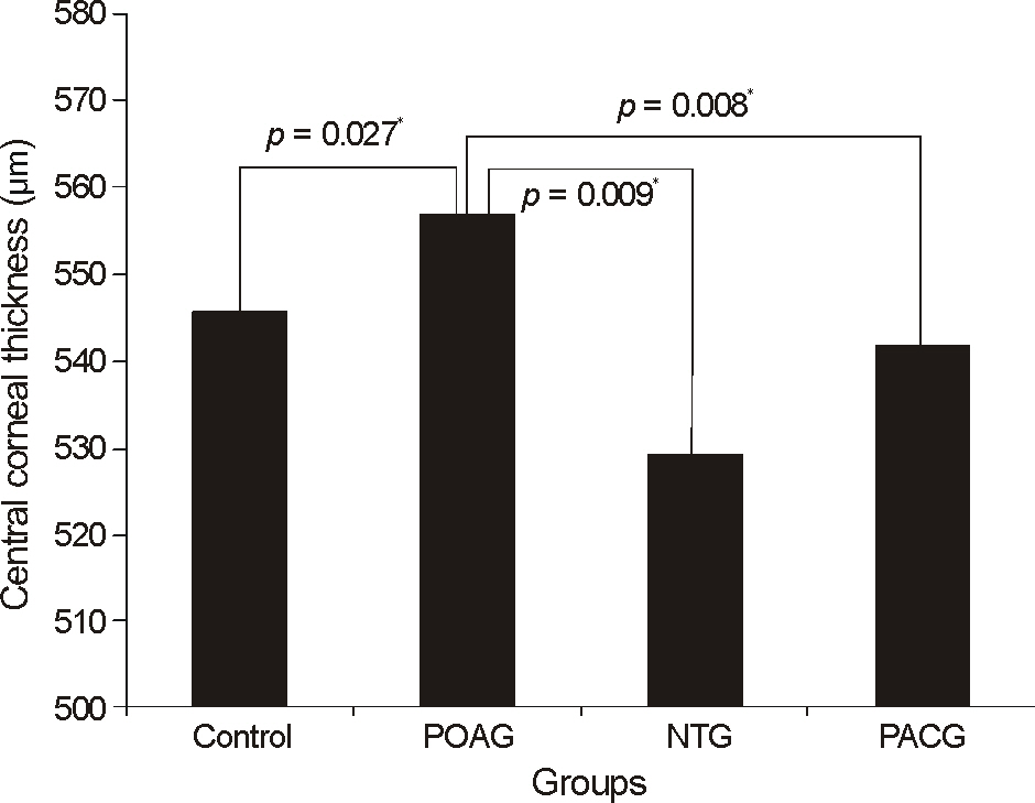
Figure 1. Comparison of central corneal thickness of all groups. *Comparison with control eyes by independent t test; *p < 0.05. POAG = primary open-angle glaucoma; HTG = high-tension glaucoma; NTG = normal tension glaucoma; PACG = primary angle-closure glaucoma.
Reference
-
References
1. Foster PJ, Buhrmann R, Quigley HA, Johnson GJ. The definition and classification of glaucoma in prevalence surveys. Br J Ophthalmol. 2002; 86:238–42.
Article2. Hu CN. An epidemiologic study of glaucoma in Shunyi County, Beijing. Zhonghua Yan Ke Za Zhi. 1989; 25:115–9.3. Wu LL, Suzuki Y, Ideta R, Araie M. Central corneal thickness of normal tension glaucoma patients in Japan. Jpn J Ophthalmol. 2000; 44:643–7.
Article4. Pang CE, Lee KY, Su DH, et al. Central corneal thickness in Chinese subjects with primary angle closure glaucoma. J Glaucoma. 2011; 20:401–4.
Article5. Seo JY, Park IW, Chung YS. Diverse types of glaucoma in patients with pseudoexfoliation syndrome: normal pressure glaucoma. J Korean Ophthalmol Soc. 2011; 52:1455–60.
Article6. Anderson DR. Automated static perimetry. St. Louis: Mosby Year Book;1992. p. 123.7. Lee ES, Kim CY, Ha SJ, et al. Central corneal thickness of Korean patients with glaucoma. Ophthalmology. 2007; 114:927–30.
Article8. Sung KR, Kim DY, Nam YP. Relationship between central corneal thickness and retinal nerve fiber layter thickness in glaucomatous subject. J Korean Ophthalmol Soc. 2009; 50:418–23.9. Copt RP, Thomas R, Mermoud A. Corneal thickness in ocular hypertension, primary open-angle glaucoma, and normal tension glaucoma. Arch Ophthalmol. 1999; 117:14–6.
Article10. Gazzard G, Foster PJ, Devereux JG, et al. Intraocular pressure and visual field loss in primary angle closure and primary open angle glaucomas. Br J Ophthalmol. 2003; 87:720–5.
Article11. Ehlers N, Hansen FK. Central corneal thickness in low-tension glaucoma. Acta Ophthalmol (Copenh). 1974; 52:740–6.
Article12. Hwang JH, Kim JS, Lee JH. Correlation between central corneal thickness and retinal nerve fiber layer thickness in normal tension glaucoma. J Korean Ophthalmol Soc. 2010; 51:63–9.
Article13. Ventura AC, Böhnke M, Mojon DS. Central corneal thickness measurements in patients with normal tension glaucoma, primary open angle glaucoma, pseudoexfoliation glaucoma, or ocular hypertension. Br J Ophthalmol. 2001; 85:792–5.
Article14. Mello PR, Meirelles SH, Moraes HV Júnior. Correlation between central corneal thickness and axial length in patients with glaucoma and normal eyes [Portuguese]. Arq Bras Oftalmol. 2009; 72:497–502.15. Xu L, Zhang H, Wang YX, Jonas JB. Central corneal thickness and glaucoma in adult Chinese: the Beijing Eye Study. J Glaucoma. 2008; 17:647–53.16. Ehlers N, Bramsen T, Sperling S. Applanation tonometer and central corneal thickness. Acta Ophthalmol. 1967; 77:734–40.17. Herndon LW, Weizer JS, Stinnett SS. Central corneal thickness as a risk factor for advanced glaucoma damage. Arch Ophthalmol. 2004; 122:17–21.
Article18. Kim JW, Chen PP. Central corneal pachymetry and visual filed progress in patients with open angle glaucoma. Ophthalmology. 2004; 111:2126–32.19. Kim JH, Lee EK, Kim CS, Lee NH. Central corneal thickness and visual field progression in primary open angle glaucoma. J Korean Ophthalmol Soc. 2007; 48:1088–95.20. Lesk MR, Hafez AS, Descovich D. Relationship between central corneal thickness and changes of optic nerve head topography and blood flow after intraocular pressure reduction in open-angle glaucoma and ocular hypertension. Arch Ophthalmol. 2006; 124:1568–72.
Article21. Henderson PA, Medeiros FA, Zangwill LM, Weinreb RN. Relationship between central corneal thickness and retinal nerve fiber layer thickness in ocular hypertensive patients. Ophthalmology. 2005; 112:251–6.
Article
- Full Text Links
-
- Actions
-
Cited
- CITED
-
- Close
- Share
-
- Similar articles
-
- The Corneal Endothelial Cell in Glaucoma and Ocular Hypertension
- Comparision of Anterior Segment Parameters in Angle-Closure Glaucoma Using Scheimpflug Camera
- Biometric Comparisons between Acute and Chronic Angle Closure Glaucoma
- Comparison of Anterior Chamber Depth with Posture Change Between Primary and lens-induced Angle Closure Glaucomas
- The Change of the Corneal Endothelial Cell after Acute Angle Closure Glaucoma
