J Korean Ophthalmol Soc.
2013 Mar;54(3):432-436. 10.3341/jkos.2013.54.3.432.
Efficacy and Intraoperative Characteristics of DisCoVisc for Cataract Surgery
- Affiliations
-
- 1Department of Ophthalmology and Visual Science, Yeouido St. Mary's Hospital, The Catholic University of Korea College of Medicine, Seoul, Korea. sara514@catholic.ac.kr
- KMID: 2216762
- DOI: http://doi.org/10.3341/jkos.2013.54.3.432
Abstract
- PURPOSE
To compare the efficacy and intraoperative characteristics of DisCoVisc with those of Hyal 2000 (sodium hyaluronate 1.0%) in cataract surgery.
METHODS
Cataract surgery was performed on 60 eyes in 49 patients who were diagnosed with moderate cataracts. 30 eyes were performed with DisCoVisc and a control group with 30 eyes using Hyal 2000 (sodium hyaluronate 1.0%). Phacodynamics was evaluated including ultrasound (US) time, mean US intensity, cumulative dissipated energy (CDE), and amount of used balanced salt solution. Corneal endothelium and corneal thickness were measured preoperatively and 1 day and 1 month and 2 months postoperatively.
RESULTS
There were no statistically significant differences in phacodynamic parameters in the two groups. The central corneal thickness change from preoperatively to postoperatively in the DisCoVisc group was +0.07 +/- 2.44 microm and Hyal 2000 group +0.84 +/- 2.93 microm (p = 0.032) at 2 months. Corneal endothelial cell loss (ECL)(%) at 2 months was 7.67 +/- 8.01% in DisCoVisc group and 13.23 +/- 15.5% in the Hyal 2000 group (p = 0.005).
CONCLUSIONS
There was signicant difference between DisCoVisc and Hyal 2000 in the changes of CCT, ECD (Endothelial cell density). (DisCoVisc is effective and provides advantages both cohesive OVDs and dispersive OVDs).
Figure
-

Figure 1. Change of preoperative and postpoerative central corneal thickness (μm). p = 0.032.
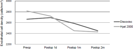
Figure 2 . Change of preoperative and postoperative corneal endothelial cell density (cells/mm2).
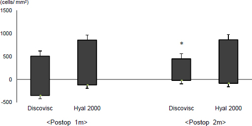
Figure 3. Endothelial cell loss between preoperation and follow up 1 m, 2 m. *Endothelial cell loss rate follow up 2 m (p = 0.005).
Reference
-
References
1. Storr-Paulsen A, Norregaard JC, Ahmed S, et al. Endothelial cell damage after cataract surgery: divide-and-conquer versus phaco-chop technique. J Cataract Refract Surg. 2008; 34:996–1000.
Article2. Fishkind W, Bakewell B, Donnenfeld ED, et al. Comparative clinical trial of ultrasound phacoemulsification with and without the WhiteStar system. J Cataract Refract Surg. 2006; 32:45–9.
Article3. Hoffman RS, Fine IH, Packer M. New phacoemulsification technology. Curr Opin Ophthalmol. 2005; 16:38–43.
Article4. Madsen K, Steveni U, Apple DJ, et al. Histochemical and receptor binding studies of hyaluronic acid binding sites on the corneal endothelium. Ophthalmic Pract. 1989; 7:92–7.5. Arshinoff SA. Dispersive-cohesive viscoelastic soft shell technique. J Cataract Refract Surg. 1999; 25:167–73.
Article6. Rainer G, Schmid KE, Findl O, et al. Natural course of intraocular pressure after cataract surgery with sodium hyaluronate 1% versus hydroxypropylmethylcellulose 2%. Ophthalmology. 2007; 114:1089–93.
Article7. Petroll WM, Jafari M, Lane SS, et al. Quantitative assessment of ophthalmic viscosurgical device retention using in vivo confocal microscopy. J Cataract Refract Surg. 2005; 31:2363–8.
Article8. Dick HB, Schwenn O. Viscoelastics in ophthalmic surgery. New York: Springer Verlag;2000. p. 7–24.9. Holzer MP, Tetz MR, Auffarth GU, et al. Effect of Healon5 and 4 other viscoelastic substances on intraocular pressure and endothelium after cataract surgery. J Cataract Refract Surg. 2001; 27:213–8.
Article10. Kim H, Joo CK. Efficacy of the soft-shell technique using Viscoat and Hyal-2000. J Cataract Refract Surg. 2004; 30:2366–70.
Article11. Binder PS, Sternberg H, Wickman MG, Worthen DM. Corneal endothelial damage associated with phacoemulsification. Am J Ophthalmol. 1976; 82:48–54.12. Petroll WM, Jafari M, Lane SS, et al. Quantitative assessment of ophthalmic viscosurgical device retention using in vivo confocal microscopy. J Cataract Refract Surg. 2005; 31:2363–8.
Article
- Full Text Links
-
- Actions
-
Cited
- CITED
-
- Close
- Share
-
- Similar articles
-
- Comparison of Balanced Salt Solution and Ophthalmic Viscosurgical Device to Maintain Optical Clarity During Phacoemulsification
- Clinical Evaluation of Traumatic Cataract with Corneal Laceration
- Comparison between Active and Gravity-based Phacoemulsification Fluidics Systems in Immediate Sequential Bilateral Cataract Surgery
- Risk Factors for Development of Posterior Capsule Opacification after Cataract Surgery or Combined Vitreoretinal Surgery
- Characteristics of Retinal Detachment in Pseudophakic Eyes
