J Korean Ophthalmol Soc.
2014 Aug;55(8):1132-1138.
Risk Factors for Development of Posterior Capsule Opacification after Cataract Surgery or Combined Vitreoretinal Surgery
- Affiliations
-
- 1Department of Ophthalmology, Maryknoll Medical Center, Busan, Korea. Pjm1438@hanmail.net
- 2Department of Ophthalmology, Haeundae Paik Hospital, Inje University College of Medicine, Busan, Korea.
Abstract
- PURPOSE
To evaluate the risk factors for the development of posterior capsule opacification (PCO) after cataract surgery or combined cataract and vitreoretinal surgery.
METHODS
In the present study all surgical procedures were performed by the same surgeon. We retrospectively reviewed 272 consecutive eyes that received cataract surgery or combined cataract and vitreoretinal surgery. The risk factors including gender, age, diabetes, continuous curvilinear capsulorhexis (CCC) size, intraocular lens shape, intraoperative intravitreal bevacizumab, gas, and silicone oil injections were evaluated using multiple logistic regression analysis.
RESULTS
PCO developed in 55 (20.2%) out of 272 eyes. The mean age was 63.3 +/- 12.1 years (range 23-85 years) and mean follow-up period was 17.3 +/- 3 months. A correlation existed between the development of the PCO and age (p < 0.05), CCC size (p = 0.009), vitreoretinal surgery (p = 0.014), intraoperative intravitreal gas (p = 0.009) and silicone oil injections (p = 0.005). However, no statistical correlation with gender, diabetes, intraocular lens shape, or intraoperative intravitreal bevacizumab injection was observed (p > 0.05).
CONCLUSIONS
The risk factors associated with PCO included young age, large CCC size, combined cataract and vitreoretinal surgery, intraoperative intravitreal gas and silicone oil injections.
MeSH Terms
Figure
-
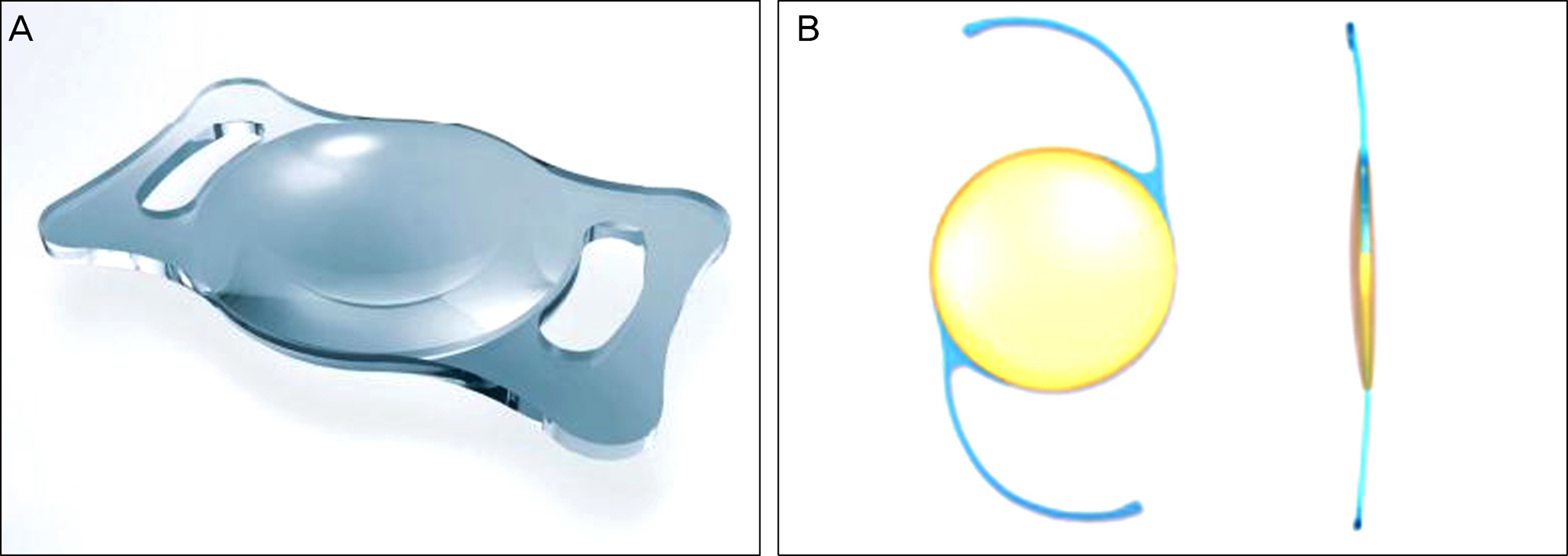
Figure 1. Photograph of two IOLs. (A) PC-60AD (HOYA®, Medical Singapore, Japan), (B) Acri. Lyc 44S (Carl Zeiss®, Meditec, Germany). IOL= intraocular lens.
Reference
-
References
1. Awasthi N, Guo S, Wagner BJ. Posterior capsular opacification; a problem reduced but not yet eradicated. Arch Ophthalmol. 2009; 127:555–62.2. Apple DJ, Solomon KD, Tetz MR, et al. Posterior capsule opacification. Surv Ophthalmol. 1992; 37:73–116.
Article3. Raj SM, Vasavada AR, Johar KS, et al. Post-operative capsular opacification: a review. Int J Biomed Sci. 2007; 3:237–50.4. Werner L, Apple DJ, Pandey SK. Postoperative proliferation of anterior and equatorial lens epithelial cells: a comparison between various foldable IOL designs. In : Buratto L, Osher RH, Masket S, editors. Cataract Surgery in Complicated Cases. Thorofare: Slack Inc.;2000. p. 399–417.5. Apple DJ, Peng Q, Vitessook N, et al. Eradication of posterior capsule opacification: documentation of a marked decrease in Nd:YAG laser posterior capsulotomy rates noted in an analysis of 5416 pseudophakic human eyes obtained postmortem. Ophthalmology. 2001; 108:505–18.6. Nishi O, Nish K, Sakka Y, et al. Intercapsular cataract surgery with lens epithelial cell removal. Part IV: Capsular fibrosis induced by poly(methymethacrylate). J Cataract Refract Surg. 1997; 17:471–7.7. Sterling S, Wood TO. Effect of intraocular lens convexity on posterior capsule opacification. J Cataract Refract Surg. 1986; 12:655–7.
Article8. Born CP, Ryan DK. Effect of intraocular lens optic design on posterior capsular opacification. J Cataract Refract Surg. 1990; 16:188–92.
Article9. Legler UFC, Apple DJ, Assia El, et al. Inhibition of posterior capsule opacification: the effect of colchicine in a sustained drug delivery system. J Cataract Refract Surg. 1993; 19:462–70.
Article10. Power WJ, Neylan D, Collum LM. Daunomycin as an inhibitor of human lens epithelial cell proliferation in culture. J Cataract Refract Surg. 1994; 20:287–90.
Article11. Lee HI, Kim MK, Ko JH, et al. Effect of polyethylene glycol polymerization onto a foldable intraocular lens in pathogenesis of posterior capsular opacity. J Korean Ophthalmol Soc. 2006; 47:621–28.12. Cho YT, Lee EH. Evaluation for the accuracy of the SRK/T Formula in PCL Implanted Patients (I). J Korean Opthalmol Soc. 1991; 32:752–60.13. Kim ER, Seo SW, Chung IY, Song JK. 2-year results of mamorylens hydrophilic acrylic intraocular lenses after catract surgery. J Korean Ophthalmol Soc. 2007; 48:356–62.14. Hollick EJ, Spalton DJ, Meacock WR. The effect of capsulorhexis size on posterior capsular opacification; one one-year results of a randomized prospective trial. Am J Ophthalmol. 1999; 128:271–9.15. Hollick EJ, Spalton DJ, Ursell PG, et al. Posterior capsular opacification with hydrogel, polymethylmethacrylate, and silicone intraocular lenses: two-year results of a randomized prospective trial. Am J Ophthalmol. 2000; 129:577–84.
Article16. Nishi O, Nishi K. Preventing posterior capsule opacification by creating a discontinuous sharp bend in the capsule. J Cataract Refract Surg. 1999; 25:521–6.
Article17. Nishi O, Nishi K, Menapace R. Capsule-bending ring for the prevention of capsular opacification: a preliminary report. Ophthalmic Surg Lasers. 1998; 29:749–53.
Article18. Nishi O, Nishi K, Sakanishi K. Inhibition of migrating lens epithelial cells at the capsular bend created by the rectangular optic edge of a posterior chamber intraocular lens. Ophthalmic Surg Lasers. 1998; 29:587–94.
Article19. Dholakia SA, Vasavada AR, Singh R. Prospective evaluation of phacoemulsification in adults younger than 50 years. J Cataract Refract Sug. 2005; 31:1327–33.
Article20. Hayashi K, Hayashi H, Nakao F, Hayashi F. Posterior capsule opacification after cataract surgery in patients with diabetes mellitus. Am J Ophthalmol. 2002; 134:10–6.
Article21. Ferguson VM, Spalton DJ. Continued breakdown of the blood aqueous barrier following cataract surgery. Br J Ophthalmol. 1992; 76:453–6.
Article22. Yoo JS, Kim MS, Lee JS. The comparison of posterior capsular opacity after cataract operation in normal and diabetic retinopathy patients. J Korean Ophthalmol Soc. 1999; 40:1860–70.23. Hollick EJ, Spalton DJ, Meacock WR. The effect of capsulorhexis size on posterior capsular opacification: one-year results of a randomized prospective trial. Am J Ophthalmol. 1999; 128:271–9.
Article24. Aykan U, Bilge AH, Karadayi K, Akin T. The effect of capsulorhexis size on development of posterior capsule opacification: small (4.5 to 5.5 mm) versus large (6.0 to 7.0 mm). Eur J Ophthalmol. 2003; 13:541–5.25. Ariki G, Ogino N. Postoperative anterior chamber inflammation after posterior chamber intraocular lens implantation concurrent with pars plana vitrectomy and lensectomy. Nippon Ganka Gakkai Zasshi. 1992; 96:1300–5.26. Sanchez-Castro GY, Hitos-Fajer A, Mendoza-Schuster , et al. Posterior capsule opacification and neovascularization treated with intravitreal bevacizumab and Nd:YAG capsulotomy. Clin Ophthalmol. 2008; 2:657–60.27. Bodanowitz S, Kir N, Hesse L. Silicone oil for recurrent vitreous hemorrhage in previously vitrectomized diabetic eyes. Ophthalmologica. 1997; 211:219–22.
Article28. Heimann K, Dahl B, Dimopoulos S, Lemmen KD. Pars plana vitrectomy and silicone oil injection in proliferative diabetic retinopathy. Graefes Arch Clin Exp Ophthalmol. 1989; 227:152–6.
Article29. Scharwey K, Pavlovic S, Jacobi KW. Early posterior capsule fibrosis after combined cataract and vitreoretinal surgery with intraocular air/SF6 gas tamponade. Klin Monbl Augenheilkd. 1998; 212:149–53.
- Full Text Links
-
- Actions
-
Cited
- CITED
-
- Close
- Share
-
- Similar articles
-
- Posterior Capsule Opacification and Intraocular Lens Design in Sulcus Fixated Posterior Chamber Lens
- A Case of Liquefied Posterior Capsular Opacification
- Posterior Capsule Opacification and Intraocular Lens Design with In-the-bag fixated Posterior Chamber Lens
- After-Cataract Following Pars Plana Lensectomy and PCL Implantation
- Posterior Chamber Intraocular Lens Implantation Combined with Pars Plana Lensectomy and Intraocular Foreign Body Removal
