A Case of Bilateral Endogenous Endophthalmitis in a Streptococcus pneumoniae Meningitis Patient
- Affiliations
-
- 1Department of Ophthalmology, Dankook University Medical College, Seoul, Korea. changmh@dankook.ac.kr
- KMID: 2216677
- DOI: http://doi.org/10.3341/jkos.2013.54.2.370
Abstract
- PURPOSE
To report a case of bilateral endogenous endophthalmitis in a Streptococcus pneumoniae meningitis patient.
CASE SUMMARY
A 45-year-old woman with bacterial meningitis was referred to the ophthalmologic clinic with acute visual impairment in both eyes. The patient's visual acuities were hand motion in both eyes. Ophthalmoscopy revealed inflammation in the anterior chamber and vitreous opacities in both eyes. Streptococcus pneumoniae was isolated in the cerebrospinal fluid sample, but not in vitreal, blood samples. Vision improved to 70/100 in the right eye and 2/100 in the left eye after 8 days of treatment.
CONCLUSIONS
Endogenous endophthalmitis constitutes a rare complication of Streptococcus pneumoniae meningitis, and a prompt diagnosis and administration of empirical intravitreal antibiotics can lead to a more favorable visual prognosis.
MeSH Terms
Figure
-
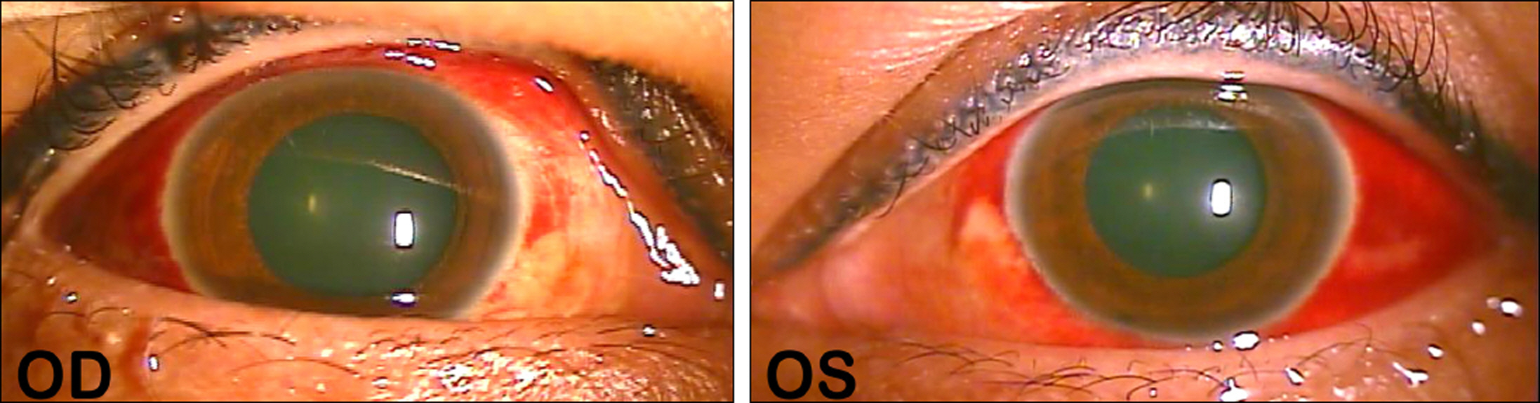
Figure 1. Slit lamp photographs at presentation. Both eye show marked conjunctival hyperemia, subconjunctival hemorrhage, and mild chemosis.
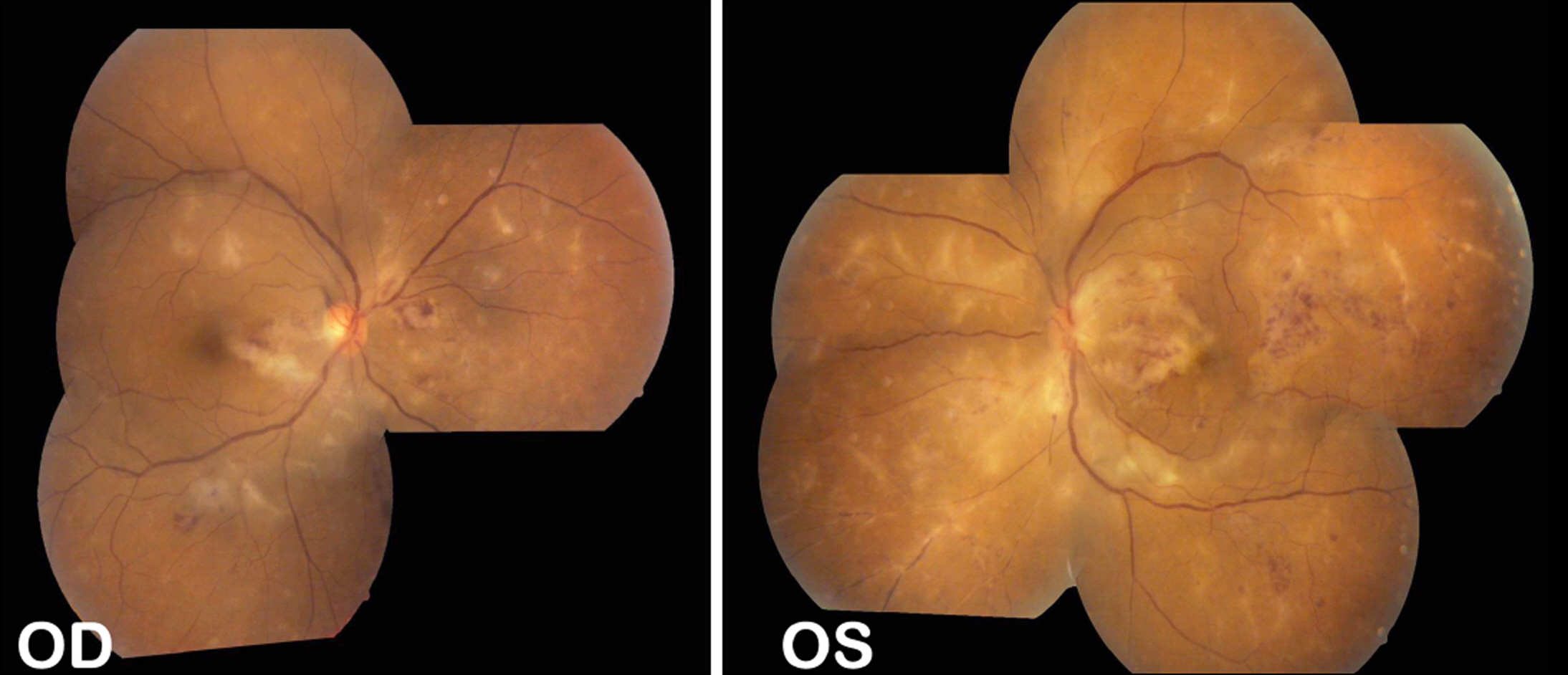
Figure 2. Fundus photographs at presentation. Right eye shows multiple ischemic changes and mild retinal hemorrhage. Left eye shows multiple ischemic changes, moderate retinal hemorrhage, and obstructed vessels.
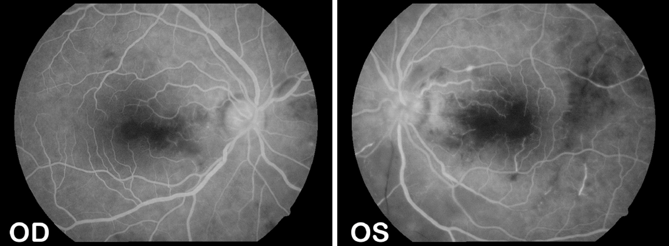
Figure 3. Fluorescein angiography at presentation. Venous phase fluorescein angiogram of both eyes demonstrating occlusive retinal vasculitis, retinal hemorrhages. Note the peripheral retina with extended leakage of inflamed vessels and optic disc.

Figure 4. Brain M R Images at presentation. Brain M RI enhance images (transverse section) show diffuse prominent leptomengeal enhancement, most suggested meningitis.
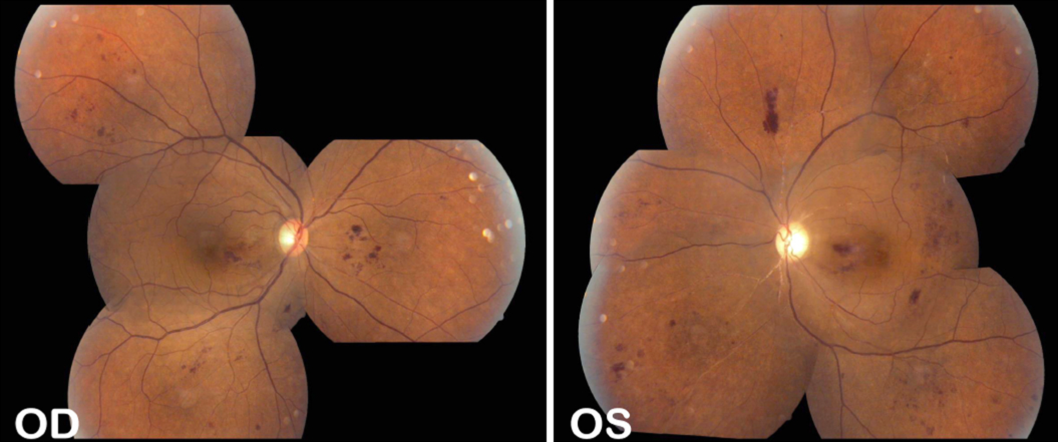
Figure 5. Fundus photographs at 6 months after treatment. Right eye shows mild retinal hemorrhage. Left eye shows mild retinal hemorrhage, ghost vessels on peripapillary area and a pale optic disc.
Reference
-
References
1. Durand ML, Calderwood SB, Weber DJ, et al. Acute bacterial meningitis in adults. A review of 493 episodes. N Engl J Med. 1993; 328:21–8.2. Miller JJ, Scott IU, Flynn HW Jr, et al. Endophthalmitis caused by Streptococcus pneumoniae. Am J Ophthalmol. 2004; 138:231–6.
Article3. Greenwald MJ, Wohl LG, Sell CH. Metastatic bacterial endophthalmitis: a contemporary reappraisal. Surv Ophthalmol. 1986; 31:81–101.
Article4. Cheesbrough JS, Williams CL, Rustom R, et al. Metastatic pneumococcal endophthalmitis: report of two cases and review of literature. J Infect. 1990; 20:231–6.
Article5. De Groot V, Stempels N, Tassingnon MJ. Endogenous pneumococcal endophthalmitis after splenectomy: report of two cases. Bull Soc Belge Ophthalmol. 1992; 243:147–51.6. Jackson TL, Eykyn SJ, Graham EM, Stanford MR. Endogenous bacterial endophthalmitis: a 17-year prospective series and review of 267 reported cases. Surv Ophthalmol. 2003; 48:403–23.
Article7. Das T, Kunimoto DY, Sharma S, et al. Relationship between clinical presentation and visual outcome in postoperative and post-traumatic endophthalmitis in south central India. Indian J Ophthalmol. 2005; 53:5–16.
Article8. Smith SR, Kroll AJ, Lou PL, Ryan EA. Endogenous bacterial and fungal endophthalmitis. Int Ophthalmol Clin. 2007; 47:173–83.
Article9. Lee SM, Lee JH. A case of Enterococcus faecalis endophthalmitis with corneal ulcer. J Korean Ophthalmol Soc. 2004; 18:175–9.
Article10. Bae JH, Lee SS. A case of Enterococcus faecalis endophthalmitis following ECCE. J Korean Ophthalmol Soc. 1994; 35:70–3.11. Kim US, Yu SY, Kwak HW. Two cases of Enterococcus fecalis endophthalmitis. J Korean Ophthalmol Soc. 2003; 44:523–8.12. Lee YH, Choi SJ, Kim IC, Chung YT. A case of the bilateral meta-static endophthalmitis. J Korean Ophthalmol Soc. 1995; 36:2048–53.13. Byun YC, Lee H, Lee EK, Lee KW. A case of metastatic endophthalmitis originated from bacterial endocardits. J Korean Ophthalmol Soc. 1994; 35:122–7.14. Lee SS, Shim HS, Park JM, Song JK. Three cases of the metastatic endophthalmitis. J Korean Ophthalmol Soc. 1994; 35:349–55.15. Han DP, Wisniewski SR, Wilson LA, et al. Spectrum and susceptibilities of microbiologic isolates in the Endophthalmitis Vitrectomy Study. Am J Ophthalmol. 1996; 122:1–17.
Article16. John JM, Ingrid US, Harry WF, et al. Endophthalmitis caused by Streptococcus pneumoniae. Am J Ophthalmol. 2004; 138:231–6.
- Full Text Links
-
- Actions
-
Cited
- CITED
-
- Close
- Share
-
- Similar articles
-
- Endogenous Streptococcus pneumoniae Endophthalmitis in Diabetic Patients
- Two Cases of Bilateral Endogenous Klebsiella pneumoniae Endophthalmitis in Primary Klebsiella pneumoniae Liver Abscess Patients
- Endogenous Pneumococcal Endophthalmitis in a Splenectomy Patient: Case Report
- Two Cases of Endogenous Endophthalmitis
- A Case of Endogenous Streptococcus Mitis Endophthalmitis in a Patient with Staphylococcus Aureus Sepsis
