J Korean Ophthalmol Soc.
2012 Aug;53(8):1172-1176. 10.3341/jkos.2012.53.8.1172.
A Case of Hydrophilic Acrylic Intraocular Lens Opacification in a Patient with Proliferative Diabetic Retinopathy
- Affiliations
-
- 1Department of Ophthalmology, Samsung Changwon Hospital, Sungkyunkwan University School of Medicine, Changwon, Korea.
- 2Department of Ophthalmology, Keimyung University Dongsan Medical Center, Keimyung University School of Medicine, Daegu, Korea. eyedr@dsmc.or.kr
- KMID: 2215963
- DOI: http://doi.org/10.3341/jkos.2012.53.8.1172
Abstract
- PURPOSE
To report a case of hydrophilic acrylic intraocular lens (IOL) opacification in a patient who underwent vitrectomy and cataract surgery for the treatment of diabetic retinopathy.
CASE SUMMARY
A 54-year-old female complained of blurred vision for 15 months after having combined vitrectomy and phacoemulsification with IOL (Rayner(R) Superflex(R) 620H) implantation for the treatment of high-risk proliferative diabetic retinopathy. On slit-lamp examination, IOL opacification was evident. IOL exchange was performed and the explanted IOL analyzed. Scanning electron microscopy (SEM) revealed the presence of translucent granular deposits on the anterior subsurface of the IOL. Energy dispersive X-ray spectroscopy (EDS) demonstrated calcium and phosphate deposition within the IOL optic.
MeSH Terms
Figure
-
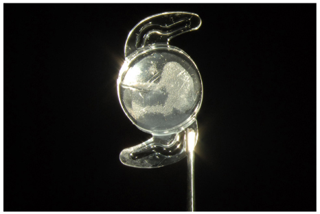
Figure 1 Slitlamp examination shows transparent granular opacification on the anterior surface of the explanted intraocular lens.
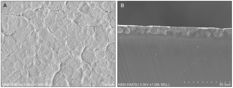
Figure 2 Scanning electron microscopic images from the explanted intraocular lens (original magnification × 1000). (A) Image of the anterior surface showing the densely packed ovoid deposits. (B) Image of cut section of the optic showing the spherical deposits beneath the anterior surface of intraocular lens.

Figure 3 The energy-dispersive X-ray analysis showed the presence of calcium and phosphate (top) within the deposits of the anterior lens surface (pink rectangle area) (bottom).
Reference
-
1. Ridley F. Safety requirements for acrylic implants. Br J Ophthalmol. 1957. 41:359–367.2. Mazzocco TR. Early clinical experience with elastic lens implants. Trans Ophthalmol Soc U K. 1985. 104(Pt 5):578–579.3. Brint SF, Ostrick DM, Bryan JE. Keratometric cylinder and visual performance following phacoemulsification and implantation with silicone small-incision or poly(methyl methacrylate) intraocular lenses. J Cataract Refract Surg. 1991. 17:32–36.4. Steinert RF, Brint SF, White SM, Fine IH. Astigmatism after small incision cataract surgery. A prospective, randomized, multicenter comparison of 4- and 6.5-mm incisions. Ophthalmology. 1991. 98:417–423.5. Levy JH, Pisacano AM, Chadwick K. Astigmatic changes after cataract surgery with 5.1 mm and 3.5 mm sutureless incisions. J Cataract Refract Surg. 1994. 20:630–633.6. Menapace R, Radax U, Amon M, Papapanos P. No-stitch, small incision cataract surgery with flexible intraocular lens implantation. J Cataract Refract Surg. 1994. 20:534–542.7. Utrata PJ, Sanders DR, DeLuca M, et al. Small incision surgery with the STAAR Elastimide three-piece posterior chamber intraocular lens. J Cataract Refract Surg. 1994. 20:426–431.8. Kohnen T. The variety of foldable intraocular lens materials. J Cataract Refract Surg. 1996. 22:Suppl 2. 1255–1258.9. Barrett G, Constable IJ. Corneal endothelial loss with new intraocular lenses. Am J Ophthalmol. 1984. 98:157–165.10. Barrett GD, Constable IJ, Stewart AD. Clinical results of hydrogel lens implantation. J Cataract Refract Surg. 1986. 12:623–631.11. Carlson KH, Cameron JD, Lindstrom RL. Assessment of the blood-aqueous barrier by fluorophotometry following poly(methyl methacrylate), silicone, and hydrogel lens implantation in rabbit eyes. J Cataract Refract Surg. 1993. 19:9–15.12. Saika S, Miyamoto T, Ohnishi Y. Histology of anterior capsule opacification with a polyHEMA/HOHEXMA hydrophilic hydrogel intraocular lens compared to poly(methyl methacrylate), silicone, and acrylic lenses. J Cataract Refract Surg. 2003. 29:1198–1203.13. Amon M, Menapace R. Cellular invasion on hydrogel and poly(methyl methacrylate) implants. An in vivo study. J Cataract Refract Surg. 1991. 17:774–779.14. Amon M, Menapace R. In vivo observation of surface precipitates of 200 consecutive hydrogel intraocular lenses. Ophthalmologica. 1992. 204:13–18.15. Bucher PJ, Büchi ER, Daicker BC. Dystrophic calcification of an implanted hydroxyethylmethacrylate intraocular lens. Arch Ophthalmol. 1995. 113:1431–1435.16. Chang BY, Davey KG, Gupta M, Hutchinson C. Late clouding of an acrylic intraocular lens following routine phacoemulsification. Eye (Lond). 1999. 13(Pt 6):807–808.17. Werner L, Apple DJ, Escobar-Gomez M, et al. Postoperative deposition of calcium on the surfaces of a hydrogel intraocular lens. Ophthalmology. 2000. 107:2179–2185.18. Frohn A, Dick HB, Augustin AJ, Grus FH. Late opacification of the foldable hydrophilic acrylic lens SC60B-OUV. Ophthalmology. 2001. 108:1999–2004.19. Werner L, Apple DJ, Kaskaloglu M, Pandey SK. Dense opacification of the optical component of a hydrophilic acrylic intraocular lens: a clinicopathological analysis of 9 explanted lenses. J Cataract Refract Surg. 2001. 27:1485–1492.20. Yu AK, Kwan KY, Chan DH, Fong DY. Clinical features of 46 eyes with calcified hydrogel intraocular lenses. J Cataract Refract Surg. 2001. 27:1596–1606.21. Lee DH, Seo Y, Joo CK. Progressive opacification of hydrophilic acrylic intraocular lenses in diabetic patients. J Cataract Refract Surg. 2002. 28:1271–1275.22. Hong JW, Roh IH, Bae SR. Opacification of hydrophilic acrylic intraocular lens. Korean J Optom Vis Sci. 2007. 6:68–71.23. Gartaganis SP, Kanellopoulou DG, Mela EK, et al. Opacification of hydrophilic acrylic intraocular lens attributable to calcification: investigation on mechanism. Am J Ophthalmol. 2008. 146:395–403.24. Neuhann IM, Neuhann TF, Szurman P, et al. Clinicopathological correlation of 3 patterns of calcification in a hydrophilic acrylic intraocular lens. J Cataract Refract Surg. 2009. 35:593–597.25. Lee CE, Kim YC, Chang SD. Opacification of the optic of an Akreos Adapt intraocular lens. Korean J Ophthalmol. 2010. 24:371–373.26. Lee SJ, Choi JH, Sun HJ, et al. Surface calcification of hydrophilic acrylic intraocular lens related to inflammatory membrane formation after combined vitrectomy and cataract surgery. J Cataract Refract Surg. 2010. 36:676–681.27. Park DH, Shin JP, Kim HK, et al. Hydrophilic acrylic intraocular lens (Akreos AO MI60) optic opacification in patients with diabetic retinopathy. Br J Ophthalmol. 2010. 94:1688–1689.28. Purbrick RM, Stavrakas P, Porooshani H, et al. Calcification of a Rayner Centerflex 570H hydrophilic acrylic intraocular lens following vitrectomy for retinal detachment: a clinicopathologic report. Eur J Ophthalmol. 2010. 20:1082–1085.29. Walker NJ, Saldanha MJ, Sharp JA, et al. Calcification of hydrophilic acrylic intraocular lenses in combined phacovitrectomy surgery. J Cataract Refract Surg. 2010. 36:1427–1431.30. Fodor M, Petrovski G, Moe MC, et al. Spectroscopic study of explanted opacified hydrophilic acrylic intraocular lenses. Acta Ophthalmol. 2011. 89:e161–e166.31. Jensen MK, Crandall AS, Mamalis N, Olson RJ. Crystallization on intraocular lens surfaces associated with the use of Healon GV. Arch Ophthalmol. 1994. 112:1037–1042.32. Olson RJ. New cases of crystalline deposits on intraocular lenses not related to any specific viscoelastic. Arch Ophthalmol. 1995. 113:1229.33. Olson RJ, Caldwell KD, Crandall AS, et al. Intraoperative crystallization on the intraocular lens surface. Am J Ophthalmol. 1998. 126:177–184.34. Neuhann IM, Kleinmann G, Apple DJ. A new classification of calcification of intraocular lenses. Ophthalmology. 2008. 115:73–79.35. Kim CJ, Choi SK. Analysis of aqueous humor calcium and phosphate form cataract eyes with and without diabetes mellitus. Korean J Ophthalmol. 2007. 21:90–94.
- Full Text Links
-
- Actions
-
Cited
- CITED
-
- Close
- Share
-
- Similar articles
-
- Clinical Characteristics of Patients with Opacification of Hydorphilic Acrylic Intraocular Lens after Cataract Surgery
- Late Opacification of a Hydrophilic Acrylic Monofocal Intraocular Lens with Hydrophobic Surface after Vitrectomy
- Clinical Features of Patients with Opacification of Hydrophilic Acrylic Intraocular Lens in Neovascular Glaucoma
- Two Cases of Intraoperative Acute Opacification of Hydrophilic Intraocular Lens
- Opacification of the Optic of an Akreos Adapt Intraocular Lens
