Subfoveal Choroidal Thickness in Fellow Eyes of Patients with Central Serous Chorioretinopathy
- Affiliations
-
- 1Department of Ophthalmology, Konkuk University Medical Center, Konkuk University School of Medicine, Seoul, Korea. eyekim@kuh.ac.kr
- KMID: 2215857
- DOI: http://doi.org/10.3341/jkos.2012.53.7.982
Abstract
- PURPOSE
To determine the relationship between subfoveal choroidal thickness of fellow eyes and choroidal vascular hyperpermeability in unilateral central serous chorioretinopathy (CSC).
METHODS
Thirty patients with unilateral CSC and 28 normal subjects underwent enhanced depth imaging spectral-domain optical coherence tomography to evaluate bilateral subfoveal choroidal thickness. The subfoveal choroidal thickness was measured from the outer RPE border to the inner sclera border. Choroidal vascular hyperpermeability was visualized with indocyanine green angiography (ICGA) and analyzed.
RESULTS
The mean subfoveal choroidal thickness in the affected eyes (439.6 +/- 136.5 microm) was significantly thicker than that in fellow eyes (340.0 +/- 103.3 microm, p = 0.002), and both showed statistically significant difference compared with normal subjects (266.5 +/- 111.5 microm, p < 0.001, p = 0.019). The subfoveal choroidal thickness of fellow eyes with choroidal vascular hyperpermeability was 370.0 +/- 176.5 microm, which differed significantly (p = 0.037) from the choroid without choroidal vascular hyperpermeability. The choroidal thickness of acute CSC was 441.6 +/- 118.6 microm, and that of chronic CSC was 454 +/- 166.5 microm, a difference that was not statistically significant (p = 0.676).
CONCLUSIONS
The subfoveal choroid with hyperpermeability was thicker than that without hyperpermeability on ICGA in the fellow eyes of patients with unilateral CSC. Enhanced depth imaging spectral-domain optical coherence tomography can indirectly evaluate the effects of choroidal hyperpermeability by noninvasively measuring the choroidal thickness.
Keyword
MeSH Terms
Figure
-
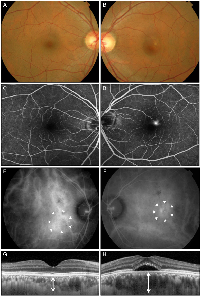
Figure 1 Representative case of a 43-year old patient with unilateral CSC. (A, B) Fundus photograph at the first examination shows serous retinal detachment of the left eye. (C, D) Juxtafoveal leaking point is seen on FA of the left eye. (E) Choroidal hyperpermeability is observed on ICGA in asymptomatic fellow eye (arrowheads). (F) Choroidal hyperpermeability surrounding leaking point on FA is also observed on ICGA (arrowheads). (G) EDI-OCT of fellow eye shows that choroidal thickness of fellow eye is thicker (305 µm, arrow) than normal. (H) EDI-OCT of affected eye shows much thickened choroid with serous detachment (481 µm).

Figure 2 Case of a 51-year-old woman with unilateral CSC. (A) Fundus photograph at the first examination reveals serous retinal detachment at the fovea. (B) FA shows the extrafoveal leakage with smokestack configuration. (C) Choroidal vascular hyperpermeability at the macula area is observed on ICGA (arrowheads). (D) No choroidal vascular hyperpermeability is observed on ICGA of the fellow eye. (E) Choroidal thickness by EDI-OCT of the affected eye is 413 µm. (F) Choroidal thickness of fellow eye without choroidal vascular hyperpermeability is thinner than that of the fellow eye with hyperpermeability. The thickness by EDI-OCT was 247 µm.
Cited by 3 articles
-
Effects of Diabetic Retinopathy and Intravitreal Bevacizumab Injection on Choroidal Thickness in Diabetic Patients
Byeong Jun Park, Hye Won Chung, Hyung Chan Kim
J Korean Ophthalmol Soc. 2013;54(10):1520-1525. doi: 10.3341/jkos.2013.54.10.1520.Influence of Image Compression on the Interpretation of Optical Coherence Tomography in Diabetic Macular Edema
Sunah Kang, Young Suk Chang, Jong Woo Kim, Chul Gu Kim, Tae Gon Lee, Jae Hui Kim
J Korean Ophthalmol Soc. 2014;55(9):1320-1326. doi: 10.3341/jkos.2014.55.9.1320.Changes in Choroidal Thickness after Panretinal Photocoagulation in Diabetic Retinopathy Patients
Sung Yu, Yong Il Kim, Kyoo Won Lee, Hyun Gu Kang
J Korean Ophthalmol Soc. 2016;57(2):256-263. doi: 10.3341/jkos.2016.57.2.256.
Reference
-
1. Bennett G. Central serous retinopathy. Br J Ophthalmol. 1955. 39:605–618.2. Negi A, Marmor MF. The resorption of subretinal fluid after diffuse damage of the retinal pigmentepithelium. Invest Ophthalmol Vis Sci. 1983. 24:1475–1479.3. Gass JD. Pathogenesis of disciform detachment of the neuroepithelium. Am J Ophthalmol. 1967. 63:Suppl. 1–139.4. Scheider A, Nasemann JE, Lund OE. Fluorescein and indocyanine green angiographies of central serous choroidopathy by scanning laser ophthalmoscopy. Am J Ophthalmol. 1993. 115:50–56.5. Guyer DR, Yannuzzi LA, Slakter JS, et al. Digital indocyanine green videoangiography of central serous chorioretinopathy. Arch Ophthalmol. 1994. 112:1057–1062.6. Prünte C. Indocyanine green angiographic findings in central serous chorioretinopathy. Int Ophthalmol. 1995. 19:77–82.7. Spaide RF, Hall L, Haas A, et al. Indocyanine green videoangiography of older patients with central serous chorioretinopathy. Retina. 1996. 16:203–213.8. Iida T, Kishi S, Hagimura N, Shimizu K. Persistent and bilateral choroidal vascular abnormalities in central serous chorioretinopathy. Retina. 1999. 19:508–512.9. Spaide RF, Koizumi H, Pozzoni MC. Enhanced depth imaging spectral-domain optical coherence tomography. Am J Ophthalmol. 2008. 146:496–500.10. Spitznas M. Pathogenesis of central serous retinopathy: a new working hypothesis. Graefes Arch Clin Exp Ophthalmol. 1986. 224:321–324.11. Gelber GS, Schatz H. Loss of vision due to central serous chorioretinopathy following psychological stress. Am J Psychiatry. 1987. 144:46–50.12. Quillen DA, Gass DM, Brod RD, et al. Central serous chorioretinopathy in women. Ophthalmology. 1996. 103:72–79.13. Scheider A, Nasemann JE, Lund OE. Fluorescein and indocyanine green angiographies of central serous choroidopathy by scanning laser ophthalmoscopy. Am J Ophthalmol. 1993. 115:50–56.14. Prünte C, Flammer J. Choroidal capillary and venous congestion in central serous chorioretinopathy. Am J Ophthalmol. 1996. 121:26–34.15. Piccolino FC, Borgia L. Central serous chorioretinopathy and indocyanine green angiography. Retina. 1994. 14:231–242.16. Piccolino FC, Borgia L, Zinicola E, Zingirian M. Indocyanine green angiographic findings in central serous chorioretinopathy. Eye (Lond). 1995. 9(Pt 3):324–332.17. Hayashi K, Hasegawa Y, Tokoro T. Indocyanine green angiography of central serous chorioretinopathy. Int Ophthalmol. 1986. 9:37–41.18. Margolis R, Spaide RF. A pilot study of enhanced depth imaging optical coherence tomography of the choroid in normal eyes. Am J Ophthalmol. 2009. 147:811–815.19. Fujiwara T, Imamura Y, Margolis R, et al. Enhanced depth imaging optical coherence tomography of the choroid in highly myopic eyes. Am J Ophthalmol. 2009. 148:445–450.20. Spaide RF. Age-related choroidal atrophy. Am J Ophthalmol. 2009. 147:801–810.21. Imamura Y, Fujiwara T, Margolis R, Spaide RF. Enhanced depth imaging optical coherence tomography of the choroid in central serous chorioretinopathy. Retina. 2009. 29:1469–1473.22. Maruko I, Iida T, Sugano Y, et al. Subfoveal choroidal thickness after treatment of central serous chorioretinopathy. Ophthalmology. 2010. 117:1792–1799.23. Kim YT, Kang SW, Bai KH. Choroidal thickness in both eyes of patients with unilaterally active central serous chorioretinopathy. Eye (Lond). 2011. 25:1635–1640.
- Full Text Links
-
- Actions
-
Cited
- CITED
-
- Close
- Share
-
- Similar articles
-
- Comparison of Choroidal Thickness in Eyes with Central Serous Chorioretinopathy, Asymptomatic Fellow Eyes and Normal Eyes
- Comparison of Intravitreal Bevacizumab and Aflibercept Injections for Central Serous Chorioretinopathy
- Comparison of Focal and Conventional Verteporfin Photodynamic Therapy for Chronic Central Serous Chorioretinopathy
- The Color Vision Test in Acute Central Serous Chorioretinopathy
- Relationships Between Choroidal Characteristics and Disease Activity in Acute Central Serous Chorioretinopathy
