Comparison of Focal and Conventional Verteporfin Photodynamic Therapy for Chronic Central Serous Chorioretinopathy
- Affiliations
-
- 1Department of Ophthalmology, KyungHee University Hospital, KyungHee University School of Medicine, Seoul, Korea. syyu@khu.ac.kr
- KMID: 2465130
- DOI: http://doi.org/10.3341/kjo.2019.0065
Abstract
- PURPOSE
To evaluate the efficacy of focal verteporfin photodynamic therapy (PDT) in patients diagnosed with chronic central serous chorioretinopathy (CSC).
METHODS
This study enrolled 52 eyes of 52 patients with chronic CSC who had received verteporfin PDT. The laser spot size of 26 eyes covering only the localized hyperfluorescent area in indocyanine green angiography was classified as focal PDT. The PDT spot size of the other 26 eyes covered the total area of retinal pigment epithelial detachment including the leaking point and was defined as conventional PDT. The central subfield thickness and subfoveal choroidal thickness were measured using Heidelberg Spectralis optical coherence tomography before PDT and at months 1, 3, 6, and 12 after PDT.
RESULTS
The mean spot size of the PDT was 1,995 µm in the focal group and 2,995 µm in the conventional group. Central subfield thickness steadily decreased in both groups. The mean baseline subfoveal choroidal thickness for the two groups was 334.95 and 348.35 µm, respectively, with no significant difference (p = 0.602). Subfoveal choroidal thickness decreased significantly to 304.20 µm at 1 month, 284.85 µm at 3 months, 271.60 µm at 6 months, and 265.95 µm at 12 months in the focal group (p < 0.001, p < 0.001, p < 0.001, and p < 0.001, respectively, compared with baseline). In the conventional group, subfoveal choroidal thickness decreased significantly to 318.75, 300, 284, and 272 µm at 1, 3, 6, and 12 months, respectively (p < 0.001, p < 0.001, p < 0.001 and p < 0.001 compared with baseline). There were no significant differences between the two groups in subfoveal choroidal thickness based on PDT spot size at 1, 3, 6, and 12 months (p = 0.633, p = 0.625, p = 0.676, and p =0.755, respectively).
CONCLUSIONS
Focal verteporfin PDT for CSC significantly decreased the subretinal fluid and sufoveal choroidal thickness to the same extent as conventional PDT.
MeSH Terms
Figure
-
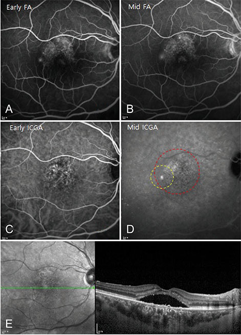
Fig. 1 Example of a chronic central serous chorioretinopathy patient who was treated with conventional photodynamic therapy (PDT). (A,B) Fluorescein angiogram before PDT showed focal leakages from the retinal pigment epithelium at the macula. Indocyanine green angiogram before PDT showed hyperfluorescence secondary to vascular hyperpermeability at the macula. (C,D) Yellow circle indicates ‘focal PDT’ area and red circle indicates ‘conventional PDT’ area in middle phase indocyanine green angiogram. (E) Spectral-domain optical coherence tomography images before PDT. FA = fluorescein angiography; ICGA = indocyanine green angiography.
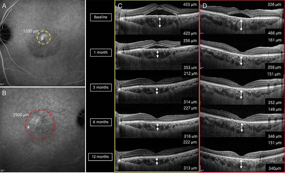
Fig. 2 (A) A case of focal photodynamic therapy (PDT) treatment in a 51-year-old male patient with chronic central subfield thickness (CSC) and (B) a case of conventional PDT treatment in a 58-year-old female patient with chronic CSC. Yellow circle indicates focal PDT area and red circle indicates conventional PDT area in middle-phase indocyanine green angiogram. Note that the focal PDT area was much smaller than the conventional area in the indocyanine green angiography. (C,D) The choroidal thickness (right bottom in optical coherence tomography) and central subfield thickness (right top in optical coherence tomography) were decreased significantly during the follow-up period compared with baseline in both groups.
Reference
-
1. Piccolino FC, Borgia L. Central serous chorioretinopathy and indocyanine green angiography. Retina. 1994; 14:231–242.2. Yannuzzi LA, Slakter JS, Gross NE, et al. Indocyanine green angiography-guided photodynamic therapy for treatment of chronic central serous chorioretinopathy: a pilot study. Retina. 2003; 23:288–298.3. Prunte C, Flammer J. Choroidal capillary and venous congestion in central serous chorioretinopathy. Am J Ophthalmol. 1996; 121:26–34.4. Castro-Correia J, Coutinho MF, Rosas V, Maia J. Long-term follow-up of central serous retinopathy in 150 patients. Doc Ophthalmol. 1992; 81:379–386.5. Levine R, Brucker AJ, Robinson F. Long-term follow-up of idiopathic central serous chorioretinopathy by fluorescein angiography. Ophthalmology. 1989; 96:854–859.6. Loo RH, Scott IU, Flynn HW Jr, et al. Factors associated with reduced visual acuity during long-term follow-up of patients with idiopathic central serous chorioretinopathy. Retina. 2002; 22:19–24.7. Cardillo Piccolino F, Eandi CM, Ventre L, et al. Photodynamic therapy for chronic central serous chorioretinopathy. Retina. 2003; 23:752–763.8. Piccolino FC, de la Longrais RR, Ravera G, et al. The foveal photoreceptor layer and visual acuity loss in central serous chorioretinopathy. Am J Ophthalmol. 2005; 139:87–99.9. Shin JY, Woo SJ, Yu HG, Park KH. Comparison of efficacy and safety between half-fluence and full-fluence photodynamic therapy for chronic central serous chorioretinopathy. Retina. 2011; 31:119–126.10. Lai TY, Chan WM, Li H, et al. Safety enhanced photodynamic therapy with half dose verteporfin for chronic central serous chorioretinopathy: a short term pilot study. Br J Ophthalmol. 2006; 90:869–874.11. Tsai MJ, Hsieh YT. Half-time photodynamic therapy for central serous chorioretinopathy. Optom Vis Sci. 2014; 91:1140–1145.12. Kang NH, Kim YT. Change in subfoveal choroidal thickness in central serous chorioretinopathy following spontaneous resolution and low-fluence photodynamic therapy. Eye (Lond). 2013; 27:387–391.13. Smretschnig E, Ansari-Shahrezaei S, Moussa S, et al. Half-fluence photodynamic therapy in acute central serous chorioretinopathy. Retina. 2012; 32:2014–2019.14. Chan WM, Lai TY, Lai RY, et al. Half-dose verteporfin photodynamic therapy for acute central serous chorioretinopathy: one-year results of a randomized controlled trial. Ophthalmology. 2008; 115:1756–1765.15. Son BK, Kim K, Kim ES, Yu SY. Long-term outcomes of full-fluence and half-fluence photodynamic therapy for chronic central serous chorioretinopathy. Ophthalmologica. 2019; 241:105–115.16. Chan WM, Lam DS, Lai TY, et al. Choroidal vascular remodelling in central serous chorioretinopathy after indocyanine green guided photodynamic therapy with verteporfin: a novel treatment at the primary disease level. Br J Ophthalmol. 2003; 87:1453–1458.17. Colucciello M. Choroidal neovascularization complicating photodynamic therapy for central serous retinopathy. Retina. 2006; 26:239–242.18. Chan WM, Lai TY, Lai RY, et al. Safety enhanced photodynamic therapy for chronic central serous chorioretinopathy: one-year results of a prospective study. Retina. 2008; 28:85–93.19. Maruko I, Iida T, Sugano Y, et al. Subfoveal choroidal thickness after treatment of central serous chorioretinopathy. Ophthalmology. 2010; 117:1792–1799.20. Pryds A, Larsen M. Choroidal thickness following extrafoveal photodynamic treatment with verteporfin in patients with central serous chorioretinopathy. Acta Ophthalmol. 2012; 90:738–743.21. Maruko I, Iida T, Sugano Y, et al. One-year choroidal thickness results after photodynamic therapy for central serous chorioretinopathy. Retina. 2011; 31:1921–1927.22. Imamura Y, Fujiwara T, Margolis R, Spaide RF. Enhanced depth imaging optical coherence tomography of the choroid in central serous chorioretinopathy. Retina. 2009; 29:1469–1473.
- Full Text Links
-
- Actions
-
Cited
- CITED
-
- Close
- Share
-
- Similar articles
-
- Chronic Central Serous Chorioretinopathy: Photodynamic Therapy
- Photodynamic Therapy With Verteporfin using Half Fluence for Chronic Central Serous Chorioretinopathy
- Photodynamic Therapy with Vertepofin for Short Time for Chronic Central Serous Chorioretinopathy
- Review and update for central serous chorioretinopathy
- The Result of Photodynamic Therapy in Chronic Central Serous Chorioretinopathy
