Glaucoma Following Pediatric Cataract Surgery: Incidence and Risk Factors
- Affiliations
-
- 1Department of Ophthalmology, Maryknoll Hospital, Busan, Korea.
- 2Department of Ophthalmology, Haeundae Paik Hospital, Inje University College of Medicine, Busan, Korea. wansookim@paik.ac.kr
- KMID: 2214996
- DOI: http://doi.org/10.3341/jkos.2011.52.10.1150
Abstract
- PURPOSE
To evaluate the incidence and risk factors of glaucoma after pediatric cataract surgery.
METHODS
We retrospectively reviewed 173 eyes which underwent pediatric cataract surgery from June 1998 to December 2009. The following parameters were ascertained: sex, laterality of cataract, age at diagnosis, age at surgery, cataract type, operation methods, optic capture, axial length (AXL), keratometry, follow-up period, and association of general abnormality.
RESULTS
Out of the 173 eyes reviewed, 8.6% were diagnosed with glaucoma. The factors not significantly different in the glaucoma group compared to the non-glaucoma group were sex, laterality of cataract, age at diagnosis, AXL, and keratometry (p > 0.05). The incidence of glaucoma was significantly higher in the aphakic group compared to the pseudophakic group. Young age at surgery, no optic capture, pars plana lensectomy, sulcus IOL implantation, and nuclear type cataract were significantly associated with increased risk of postoperative glaucoma (p < 0.05).
CONCLUSIONS
Patients with several predictors of postoperative glaucoma which may affect visual acuity may require extensive postoperative care after pediatric cataract surgery.
Keyword
MeSH Terms
Figure
-

Figure 1. Age at cataract surgery in children. Blue bar represent children who developed postcataract surgery glaucoma, and green bar represent children who did not develop postcataract surgery glaucoma.

Figure 2. Kaplan-Meier plots of glaucoma free survival by aphakic and phakic eyes. The rates of postoperative glaucoma were similar in the 2 groups for the first 50 months after surgery but thereafter, the rate of glaucoma was higher in aphakic eyes. The log rank test revealed strong evidence of association between aphakic and phakic eyes (p = 0.03).

Figure 3. This graph shows change of postoperative intraocular pressure (IOP) between aphakic and pseudophakic eyes. The difference of postoperative 1 month IOP was statistically significant between aphakic and pseudophakic eyes (p = 0.008).
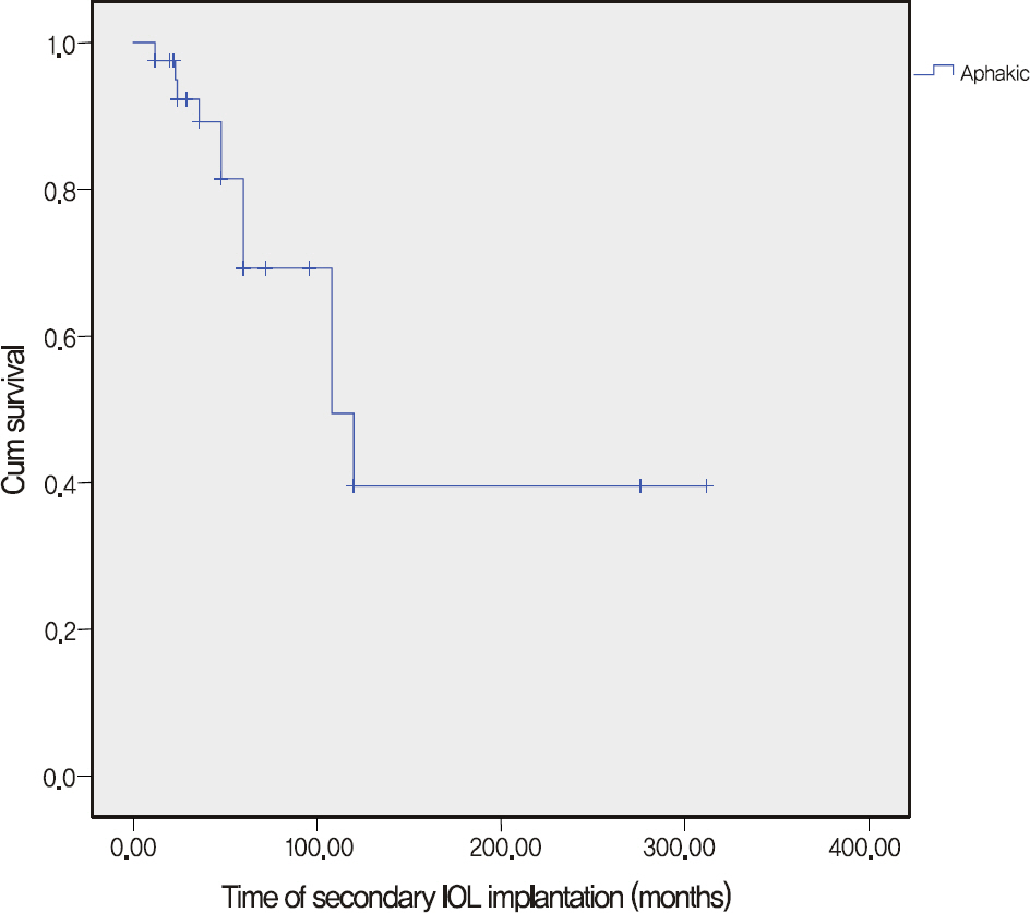
Figure 4. This graph shows higher rates of glaucoma when secondary IOL implantation was delayed, however, the rate of glaucoma did not change after 108 months.

Figure 5. Surgical technique used for pediatric cataract extraction and rate of postoperative glaucoma for each surgical technique. SD = standard deviation; PPL = Pars plana lensec-tomy; PCCC = posterior continuous curvilinear capsulorhexis. * Statistically significant (p < 0.05).
Reference
-
References
1. Steinert RF. Cataract Surgery: Technique, Complications and Management. 2nd ed.Philadelphia: WB Sauders;2004. p. 273.2. Mori M, Keech RV, Scott WE. Glaucoma and ocular hypertension in pediatric patients with cataracts. J AAPOS. 1997; 1:98–101.
Article3. Mills MD, Robb RM. Glaucoma following childhood cataract surgery. J Pediatr Ophthalmol Strabismus. 1994; 31:355–60.4. Tatham A, Odedra N, Tayebjee S, et al. The incidence of glaucoma following paediatric cataract surgery: a 20-year retrospective study. Eye (Lond). 2010; 24:1366–75.
Article5. Kirwan C, Lanigan B, O'Keefe M. Glaucoma in aphakic and pseudophakic eyes following surgery for congenital cataract in the first year of life. Acta Ophthalmol. 2010; 88:53–9.
Article6. Rabiah PK. Frequency and predictors of glaucoma after pediatric cataract surgery. Am J Ophthalmol. 2004; 137:30–7.
Article7. Ahn JH, Kim WS. Surgical techniques and postoperative complications in pediatric cataract surgery. J Korean Ophthalmol Soc. 2006; 47:1049–56.8. Swamy BN, Billson F, Martin F, et al. Secondary glaucoma after paediatric cataract surgery. Br J Ophthalmol. 2007; 91:1627–30.
Article9. Wilson ME Jr, Bartholomew LR, Trivedi RH. Pediatric cataract surgery and intraocular lens implantation: practice styles and preferences of the 2001 ASCRS and AAPOS memberships. J Cataract Refract Surg. 2003; 29:1811–20.10. Walton DS. Pediatric aphakic glaucoma: a study of 65 patients. Trans Am Ophthalmol Soc. 1995; 93:403–13.11. Birch EE, Stager DR. The critical period for surgical treatment of dense congenital unilateral cataract. Invest Ophthalmol Vis Sci. 1996; 37:1532–8.
Article12. Trivedi RH, Wilson ME Jr, Golub RL. Incidence and risk factors for glaucoma after pediatric cataract surgery with and without intraocular lens implantation. J AAPOS. 2006; 10:117–23.
Article13. Lambert SR, Lynn M, Drews-Botsch C, et al. Intraocular lens implantation during infancy: perceptions of parents and the American Association for Pediatric Ophthalmology and Strabismus members. J AAPOS. 2003; 7:400–5.
Article14. Asrani S, Freedman S, Hasselblad V, et al. Does primary intraocular lens implantation prevent “aphakic” glaucoma in children? J AAPOS. 2000; 4:33–9.
Article15. Kim DH, Kim JH, Kim SJ, Yu YS. Long-term results of bilateral congenital cataract treated with early cataract surgery, aphakic glasses and secondary IOL implantation. Acta Ophthalmol. 2010 Sep 2. [Epub ahead of print].
Article16. Scheie HG. Aspiration of congenital or soft cataracts: a new technique. Am J Ophthalmol. 1960; 50:1048–56.
Article17. Michaelides M, Bunce C, Adams GG. Glaucoma following congenital cataract surgery–the role of early surgery and posterior capsulotomy. BMC Ophthalmol. 2007; 7:13.
Article18. Gimbel HV. Posterior capsulorhexis with optic capture in pediatric cataract and intraocular lens surgery. Ophthalmology. 1996; 103:1871–5.
Article19. Gimbel HV, DeBroff BM. Posterior capsulorhexis with optic capture: maintaining a clear visual axis after pediatric cataract surgery. J Cataract Refract Surg. 1994; 20:658–64.
Article20. LeBoyer RM, Werner L, Snyder ME, et al. Acute haptic-induced ciliary sulcus irritation associated with single-piece AcrySof intraocular lenses. J Cataract Refract Surg. 2005; 31:1421–7.
Article21. Wintle R, Austin M. Pigment dispersion with elevated intraocular pressure after AcrySof intraocular lens implantation in the ciliary sulcus. J Cataract Refract Surg. 2001; 27:642–4.
Article22. Parks MM, Johnson DA, Reed GW. Long-term visual results and complications in children with aphakia. A function of cataract type. Ophthalmology. 1993; 100:826–40.23. Chen TC, Bhatia LS, Halpern EF, Walton DS. Risk factors for the development of aphakic glaucoma after congenital cataract surgery. Trans Am Ophthalmol Soc. 2006; 104:241–51.
Article
