Comparison of Corneal Higher-Order Aberrations Measured with Two Instruments Using Scheimpflug Camera System
- Affiliations
-
- 1Department of Ophthalmology, Ewha Womans University School of Medicine, Seoul, Korea. jrmoph@ewha.ac.kr
- 2BE Woorieyes Clinic, Chuncheon, Korea.
- KMID: 2214407
- DOI: http://doi.org/10.3341/jkos.2015.56.10.1497
Abstract
- PURPOSE
To compare the corneal higher-order aberrations (HOAs) of normal young subjects using Galilei(TM) G4 (Zeimer, Port, Switzerland) and Pentacam(R) (Oculus Inc., Wetzlar, Germany).
METHODS
Corneal HOAs were measured using Galilei(TM) G4 and Pentacam(R) in 41 healthy individuals (41 eyes). Intraclass correlation coefficients (ICCs) were obtained to evaluate the repeatability of the 2 devices. Differences in HOAs between the 2 instruments were analyzed with a paired t-test and correlations evaluated.
RESULTS
All ICCs measured using Galilei(TM) G4 and Pentacam(R) showed more than moderate repeatability (>0.81) except trefoil, tetrafoil, 4th and 5th HOAs. When comparing the measurements obtained with Galilei(TM) G4 and Pentacam(R), total HOAs, spherical aberration (SA), secondary astigmatism and 5th total HOAs were statistically significantly different between the 2 device (all p < or = 0.001). In addition, Galilei(TM) G4 and Pentacam(R) showed discrepancy among all corneal HOAs items. Although the total corneal HOAs and the SA were significantly correlated, other HOA measurements generally exhibited a low correlation.
CONCLUSIONS
Corneal HOAs obtained by the 2 instruments cannot be used interchangeably due to their differences and discrepancy although corneal HOAs measured using Galilei(TM) G4 and Pentacam(R) showed relatively high repeatability.
MeSH Terms
Figure
-

Figure 1. Bland-Altman plots showing the agreement between mean corneal aberrometers (μ m) obtained by the 2 tomography de-vices: Galilei TM G4 and Pentacam®. (A-F) graphs plot the difference against the average of two measurements of corneal HOAs, with 95% limits of agreement (broken lines) and the mean difference (black line). (A) Total corneal HOAs, (B) Coma, (C) Trefoil, (D) Spherical aberration, (E) Secondary astigmatism, (F) Tetrafoil. HOAs = higher-order aberrations; SA = spherical aberration; 2° astig = secondary astigmatism.
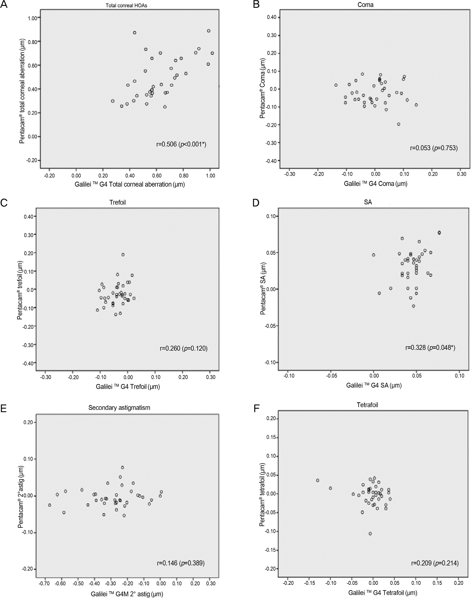
Figure 2. Correlations of measurements of corneal aberration using Galilei TM G4 and Pentacam®. (A-F) Shows measurements of total corneal HOAs using Galilei TM G4 against Pentacam® measurements. (A) Total corneal HOAs, (B) Coma, (C) Trefoil, (D) Spherical aberration, (E) Secondary astigmatism, (F) Tetrafoil. HOAs = higher-order aberrations; SA = spherical aberration; 2° astig = sec-ondary astigmatism; r = Pearson correlation coefficient. * Statistically significant ( p-value < 0.05, based on t-test).
Cited by 2 articles
-
Comparison of Anterior Segment Measurements between Dual and Single Scheimpflug Camera
Youngju An, Hyojin Kim, Choun-Ki Joo
J Korean Ophthalmol Soc. 2016;57(7):1056-1062. doi: 10.3341/jkos.2016.57.7.1056.Comparison of Corneal Astigmatism and Higher-order Aberrations between Color Light-emitting Diode Topographer and Scheimpflug Imager
Da Yeong Kim, Minji Ha, Rowoon Yi, Hyo Won Kim, So-Hyang Chung
J Korean Ophthalmol Soc. 2019;60(10):922-928. doi: 10.3341/jkos.2019.60.10.922.
Reference
-
References
1. Lombardo M, Lombardo G. Wave aberration of human eyes and new descriptors of image optical quality and visual performance. J Cataract Refract Surg. 2010; 36:313–31.
Article2. Artal P, Guirao A, Berrio E, Williams DR. Compensation of cor-neal aberrations by the internal optics in the human eye. J Vis. 2001; 1:1–8.
Article3. Campbell CE. A new method for describing the aberrations of the eye using Zernike polynomials. Optom Vis Sci. 2003; 80:79–83.
Article4. Barkana Y, Gerber Y, Elbaz U. . Central corneal thickness measurement with the Pentacam Scheimpflug system, optical low- coherence reflectometry pachymeter, and ultrasound pachymetry. J Cataract Refract Surg. 2005; 31:1729–35.5. Lee YE, Jun RM. The intra and inter-examiner repeatability of cor-neal parameters obtained by GALILEI(TM) in normal subjects. J Korean Ophthalmol Soc. 2009; 50:1611–16.6. Burakgazi AZ, Tinio B, Bababyan A. . Higher order aberra-tions in normal eyes measured with three different aberrometers. J Refract Surg. 2006; 22:898–903.
Article7. Yum JH, Choi SK, Kim JH, Lee DH. Comparison of aberrations in Korean normal eyes measured with two different aberrometers. J Korean Ophthalmol Soc. 2009; 50:1789–94.
Article8. Knapp S, Awwad ST, Ghali C, McCulley JP. Ocular aberrations measured by the Fourier-based WaveScan and Zernike-based LADARWave Hartmann-Shack aberrometers. J Refract Surg. 2009; 25:201–9.
Article9. Shin JY, Lee MY, Chung SH. Comparison of keratometry and cor-neal higher order aberrations between Scout videokeratoscope and Pentacam Scheimpflug camera. J Korean Ophthalmol Soc. 2014; 55:1758–64.
Article10. McGraw KO, Wong SP. Forming inferences about some intraclass correlation coefficients. Psychological Methods. 1996; 1:30.
Article11. Williams D, Yoon GY, Porter J. . Visual benefit of correcting higher order aberrations of the eye. J Refract Surg. 2000; 16:S554–9.
Article12. Artal P, Berrio E, Guirao A, Piers P. Contribution of the cornea and internal surfaces to the change of ocular aberrations with age. J Opt Soc Am A Opt Image Sci Vis. 2002; 19:137–43.
Article13. López-Miguel A, Maldonado MJ, Belzunce A. . Precision of a commercial hartmann-shack aberrometer: limits of total wavefront laser vision correction. Am J Ophthalmol. 2012; 154:799–807.e5.
Article14. Aramberri J, Araiz L, Garcia A. . Dual versus single Scheimpflug camera for anterior segment analysis: precision and agreement. J Cataract Refract Surg. 2012; 38:1934–49.
Article15. Cerviño A, Dominguez-Vicent A, Ferrer-Blasco Blasco. . Intrasubject repeatability of corneal power, thickness, and wavefront aberra-tions with a new version of a dual rotating Scheimpflug-Placido system. J Cataract Refract Surg. 2015; 41:186–92.
Article16. Wang L, Shirayama M, Koch DD. Repeatability of corneal power and wavefront aberration measurements with a dual-Scheimpflug Placido corneal topographer. J Cataract Refract Surg. 2010; 36:425–30.
Article17. Netto MV, Ambrósio R Jr, Shen TT, Wilson SE. Wavefront analy-sis in normal refractive surgery candidates. J Refract Surg. 2005; 21:332–8.
Article18. Yoon G, Macrae S, Williams DR, Cox IG. Causes of spherical aberration induced by laser refractive surgery. J Cataract Refract Surg. 2005; 31:127–35.
Article19. Domínguez-Vicent A, Monsálvez-Romín D, Aguila-Carrasco Carrasco. . Measurements of anterior chamber depth, white-to-white dis-tance, anterior chamber angle, and pupil diameter using two Scheimpflug imaging devices. Arq Bras Oftalmol. 2014; 77:233–7.
Article20. Al-Sayyari TM, Fawzy SM, Al-Saleh AA. Corneal spherical aber-ration in Saudi population. Saudi J Ophthalmol. 2014; 28:207–13.
Article
- Full Text Links
-
- Actions
-
Cited
- CITED
-
- Close
- Share
-
- Similar articles
-
- Comparison of Keratometry and Corneal Higher Order Aberrations between Scout Videokeratoscope and Pentacam Scheimpflug Camera
- Central Corneal Thickness Measured by Noncontact Specular Microscopy, Dual Rotating Scheimpflug Camera and Ultrasound Pachymetry
- Corneal Thickness Measured by Dual Scheimpflug, Anterior Segment Optical Coherence Tomography, and Ultrasound Pachymetry
- Comparison of Corneal Higher-order Aberrations Measured by Scheimpflug Camera and Placido Disc-based Topography in Korean Patients
- Comparison of Corneal Astigmatism and Higher-order Aberrations between Color Light-emitting Diode Topographer and Scheimpflug Imager
