J Korean Ophthalmol Soc.
2010 Jun;51(6):816-821. 10.3341/jkos.2010.51.6.816.
Anterior and Posterior Corneal Spherical Aberration Measured With Pentacam in the Korean
- Affiliations
-
- 1HanGil Eye Hospital, Korea. chobjn@empal.com
- KMID: 2213605
- DOI: http://doi.org/10.3341/jkos.2010.51.6.816
Abstract
- PURPOSE
To evaluate the spherical aberrations of the anterior and posterior surfaces of normal corneas using Pentacam in a Korean sample population and determine their ranges and changes with age.
METHODS
We used Pentacam (Oculus Inc.,Germany) to measure the anterior and posterior corneal spherical aberrations of 240 eyes in 240 patients with normal corneas who visited our clinic. The means and ranges of spherical aberrations and their changes with age were determined. We examined both eyes of 90 patients to confirm the inter-ocular symmetry in spherical aberration.
RESULTS
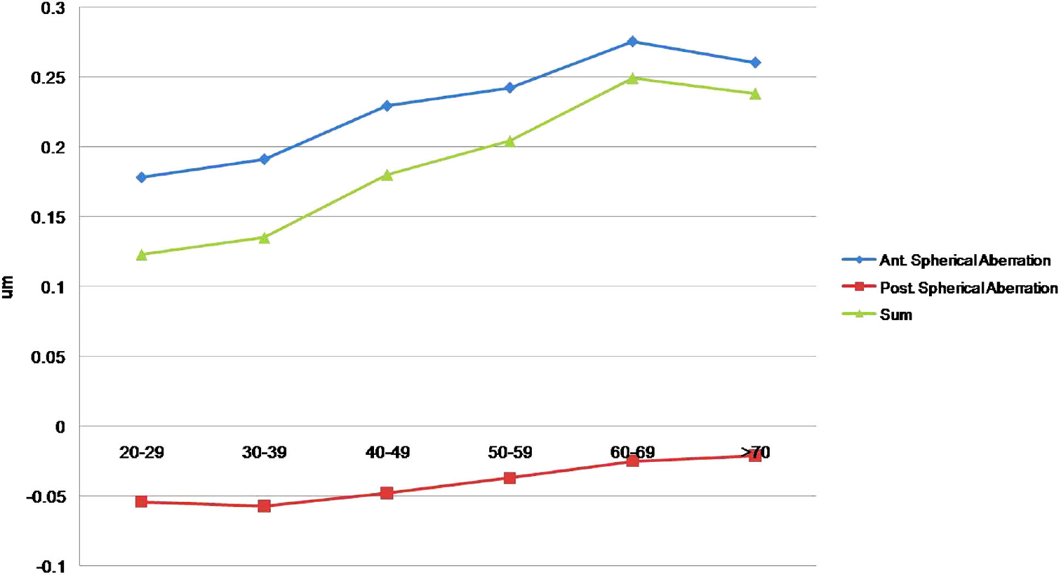
The mean age of the 240 patients (M:F=103:137) was 49.8 years (range: 20-79), and the mean spherical aberrations of the anterior and posterior surfaces of the cornea were 0.230+/-0.078 micrometer, and -0.04+/-0.021 micrometer, respectively. The mean total corneal spherical aberration was 0.19+/-0.087 micrometer. There were no differences between males and females, and inter-ocular symmetry was observed in all tested patients. There was a tendency for the values of anterior, posterior and total corneal spherical aberration to increase with age. Ranges of spherical aberrations were from -0.177 micrometer to 0.423 micrometer in the anterior cornea, from -0.083 micrometer to 0.034 micrometer in the posterior cornea, and from -0.238 micrometer to 0.410 micrometer in the total cornea.
CONCLUSIONS
In a Korean population, the mean total corneal spherical aberration was 0.19 micrometer, which was shown to increase with age. Some patients were shown to have an extreme value. Based on these results, a preoperative analysis for corneal spherical aberration may be helpful when selecting aspheric intraocular lenses.
Keyword
Figure
Cited by 2 articles
-
Comparisons of Clinical Results after Implantation of Three Aspheric Intraocular Lenses
Young Joon Choi, Kyung Eun Han, Ji Min Ahn, Se Hwan Jeong, Hyung Keun Lee, Kyoung Yul Seo, Eung Kweon Kim, Tae Im Kim
J Korean Ophthalmol Soc. 2013;54(2):251-256. doi: 10.3341/jkos.2013.54.2.251.Age-related Changes in Anterior, Posterior Corneal Astigmatism in a Korean Population
Yoon Seob Sim, Soon Won Yang, Yu Li Park, Kyung Sun Na, Hyun Seung Kim
J Korean Ophthalmol Soc. 2017;58(8):911-915. doi: 10.3341/jkos.2017.58.8.911.
Reference
-
References
1. Marshall J, Cionni RJ, Davison J, et al. Clinical results of the blue-light filtering AcrySof Natural foldable acrylic intraocular lens. J Cataract Refract Surg. 2005; 31:2319–23.
Article2. Kim HS, Kim SW, Ha BJ, et al. Ocular aberrations and contrast sensitivity in eyes implantes with aspheric and spherical intraocular lenses. J Korean Ophthalmol Soc. 2008; 49:1256–62.3. Wang L, Dai E, Koch DD, Nathoo A. Optical aberrations of the human cornea. J Caract Refract Surg. 2003; 29:1514–21.4. Bellucci R, Scialdone A, Buratto L, et al. Visual acuity and contrast sensitivity comparison between Tecnis and AcrySof SA60AT intraocular lenses: A multicenter randomized study. J Cataract Refract Surg. 2005; 31:712–7.
Article5. Packer M, Fine IH, Hoffman RS, Piers PA. Prospective randomized trial of an anterior surface modified prolate intraocular lens. J Refract Surg. 2002; 18:692–6.
Article6. Rocha K, Soriano E, Chalita M, et al. Wavefront analysis and contrast sensitivity of aspheric and spherical intraocular lenses. Am J Ophthalmol. 2006; 142:750–6.7. Tzelikis P, Akaishi L, Trindade F, Boteon J. Ocular aberrations and contrast sensitivity after cataract surgery with AcrySof IQ intraocular lens implantation. J Cataract Refract Surg. 2007; 33:1918–24.
Article8. Caporossi A, Martone G, Casprini F, Rapisarda L. Prospective randomized study of clinical performance of 3 aspheric and 2 spherical intraocular lenses in 250 eyes. J Refract Surg. 2007; 23:639–48.
Article9. Awwad ST, Lehmann JD, McCulley JP, Bowman RW. A comparison of higher order aberrations in eyes implanted with AcrySof IQ SN60WF and AcrySof SN60AT intraocular lenses. Eur J Ophthalmol. 2007; 17:320–6.
Article10. Dubbelman M, Sicam VADP, Van der Heijdg GL. The shape of the anterior and posterior surface of the aging human cornea. Vision Res. 2006; 46:993–1001.
Article11. Chen M, Yoon G. Posterior corneal aberrations and their compensation effects on anterior corneal aberrations in keratoconic eyes. Invest Ophthalmol Vis Sci. 2008; 49:5645–52.
Article12. Miranda MA, Radhakrishnan H, O'Donnell C. Repeatability of oculus pentacam metrics derived from corenal topography. Cornea. 2009; 28:657–66.13. Swaartz T, Marten L, Wang M. Measuring the cornea: the latest developments in corneal topography. Curr Opin Ophthalmol. 2007; 18:325–33.14. Mester U, Dillinger P, Anterist N. Impact of a modified optic design on visual function: clinical comparative study. J Cataract Refract Surg. 2003; 29:652–60.
Article15. Lim KL, Fam HB. Ethnic differences in higher-order aberrations: Spherical aberration in the South East Asian Chinese eye. J Cataract Refract Surg. 2009; 35:2144–8.
Article16. Amano S, Amano Y, Yamagami S, et al. Age-related changes in corneal and ocular higher-order wavefront aberrations. Am J Ophthalmol. 2004; 137:988–92.
Article17. Sicam VA, Dubbelman M, van der Heijde RG. Spherical aberration of the anterior and posterior surfaces of the human cornea. J Opt Soc Am A Opt Image Sci Vis. 2006; 23:544–9.
Article18. Guirao A, Redondo M, Artal P. Optical aberrations of the human cornea as a function of age. J Opt Soc Am A Opt Image Sci Vis. 2000; 17:1697–702.
Article19. Holladay JT, Piers PA, Koranyi G, et al. A new intraocular lens design to reduce spherical aberration of pseudophakic eyes. J Refract Surg. 2002; 18:683–91.
Article20. Beiko GH, Haigis W, Steinmueller A. Distribution of corneal spherical aberration in a comprehensive ophthalmology practice and whether keratometry can predict aberration values. J Cataract Refract Surg. 2007; 33:848–58.
Article
- Full Text Links
- Actions
-
Cited
- CITED
-
- Close
- Share
- Similar articles
-
- Comparison between Anterior Corneal Aberration and Ocular Aberration in Laser Refractive Surgery
- Changes of the Corneal Aberration Following Cataract Surgery
- Comparison of Keratometry and Corneal Higher Order Aberrations between Scout Videokeratoscope and Pentacam Scheimpflug Camera
- Changes in Higher-order Aberrations after Superior-incision Cataract Surgery in Patients with Positive Vertical Coma
- Induced Astigmatism and High-Order Aberrations after 1.8-mm, 2.2-mm and 3.0-mm Coaxial Phacoemulsification Incisions


