J Korean Ophthalmol Soc.
2011 Apr;52(4):407-413.
Induced Astigmatism and High-Order Aberrations after 1.8-mm, 2.2-mm and 3.0-mm Coaxial Phacoemulsification Incisions
- Affiliations
-
- 1Sungmo Eye Hospital, Busan, Korea. sungmo@sungmo.co.kr
Abstract
- PURPOSE
To study theeffect of micro incision (1.8 mm) and small incision (2.2 mm and 2.8 mm) coaxial phacoemulsification on surgically induced astigmatism (SIA) and high-order aberrations (HOA) of anterior and posterior corneal surface.
METHODS
The present randomized clinical study included 32 eyes having a 1.8-mm, 38 eyes having a 2.2-mm, and 30 eyes having a 2.8-mm corneal incision. SIAs were measured at 1 and 3 months postoperatively. HOAs included coma, trefoil, and spherical aberration. The coma-root mean square (RMS) and trefoil-RMS were evaluated at 1 month after the cataract operation.
RESULTS
Surgically induced astigmatisms were 0.41 +/- 0.30 diopter (D) in the 1.8-mm incision group, 0.47 +/- 0.21 D in 2.2-mm group and 0.71 +/- 0.50 D in the 2.8-mm group. The SIA of the 1.8-mm group was smaller than the other groups (p = 0.002). There was no statistically significant difference in coma, spherical aberration of the corneal anterior surface and trefoil, or spherical aberration of the posterior surface among the 3 groups at 1 month after surgery.
CONCLUSIONS
Incision size contributes to postoperative corneal astigmatism. Phacoemulsification cataract surgery with less than 2.8-mm incision does not significantly influence the corneal aberrationsof anterior and posterior corneal surfaces.
Keyword
Figure
-

Figure 1. Inter-group comparison of surgically induced astigmatism (SIA) at 1 and 3 months after cataract surgery. SIA is statistically different among 1.8-mm, 2.2-mm and 2.8-mm incision group at 1 and 3 months respectively.
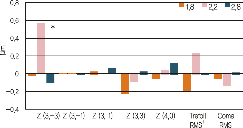
Figure 2. Mean changes (μ m) in high order aberrations and root mean square of anterior corneal surface betweenbaseline and 1 month after operation. Only Z (3, -3) is different in statistical comparison among 3 groups. *p<0.05; † root mean square.
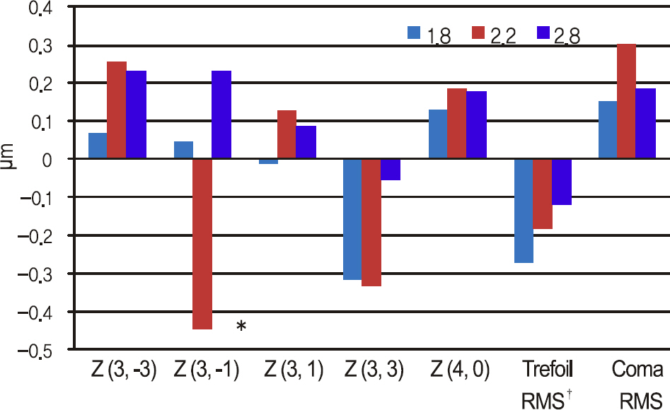
Figure 3. Mean changes (μ m) in high order aberrations and root mean square of posterior corneal surface betweenbaseline and 1 month after operation. Only Z (3, -1) is different in statistical comparison among 3 groups. *p<0.05; † root mean square.
Reference
-
References
1. Shepherd JR. Induced astigmatism in small incision cataract surgery. J Cataract Refract Surg. 1989; 15:85–8.
Article2. Steinert RF, Brint SF, White SM, Fine IH. Astigmatism after small incision cataract surgery. A prospective, randomized, multicenter comparison of 4- and 6.5-mm incisions. Ophthalmology. 1991; 98:417–23. discussion 423-4.3. Soscia W, Howard JG, Olson RJ. Microphacoemulsification with WhiteStar. A wound-temperature study. J Cataract Refract Surg. 2002; 28:1044–6.4. Donnenfeld ED, Olson RJ, Solomon R, et al. Efficacy and wound-temperature gradient of whitestar phacoemulsification through a 1.2 mm incision. J Cataract Refract Surg. 2003; 29:1097–100.
Article5. Dosso AA, Cottet L, Burgener ND, Di Nardo S. Outcomes of coaxial microincision cataract surgery versus conventional coaxial cataract surgery. J Cataract Refract Surg. 2008; 34:284–8.
Article6. Kurz S, Krummenauer F, Gabriel P, et al. Biaxial microincision versus coaxial small-incision clear cornea cataract surgery. Ophthalmology. 2006; 113:1818–26.
Article7. Elkady B, Alió JL, Ortiz D, Montalbán R. Corneal aberrations after microincision cataract surgery. J Cataract Refract Surg. 2008; 34:40–5.
Article8. Denoyer A, Denoyer L, Marotte D, et al. Intraindividual comparative study of corneal and ocular wavefront aberrations after biaxial microincision versus coaxial small-incision cataract surgery. Br J Ophthalmol. 2008; 92:1679–84.
Article9. Lee AG, Greenlee E, Oetting TA, et al. Assessing cataract surgical competency. Ophthalmology. 2007; 114:1415–6.
Article10. Osher RH. Microcoaxial phacoemulsification Part 2: clinical study. J Cataract Refract Surg. 2007; 33:408–12.11. Osher RH, Injev VP. Microcoaxial phacoemulsification Part 1: lab-oratory studies. J Cataract Refract Surg. 2007; 33:401–7.12. Tong N, He JC, Lu F, et al. Changes in corneal wavefront aberrations in microincision and small-incision cataract surgery. J Cataract Refract Surg. 2008; 34:2085–90.
Article13. Wang J, Tang X, Zhang S, Li LH. Changes in high order aberrations of anterior and posterior surfaces of cornea before and after phacoemulsification. Zhonghua Yan Ke Za Zhi. 2008; 44:1066–71.14. Lee KM, Kwon HG, Joo CK. Microcoaxial cataract surgery outcomes: comparison of 1.8 mm system and 2.2 mm system. J Cataract Refract Surg. 2009; 35:874–80.
Article15. Lee SY, Chung JL, Hong JP, et al. Comparative study of two aspheric, aberration-free intraocular lenses in cataract surgery. J Korean Ophthalmol Soc. 2009; 50:1520–6.
Article16. Hwang SJ, Choi SK, Oh SH, et al. Surgically induced astigmatism and corneal higher order aberrations in microcoaxial and conventional cataract surgery. J Korean Ophthalmol Soc. 2008; 49:1597–602.
Article17. Holladay JT, Moran JR, Kezirian GM. Analysis of aggregate surgically induced refractive change, prediction error, and intraocular astigmatism. J Cataract Refract Surg. 2001; 27:61–79.
Article18. Alió J, Rodríguez-Prats JL, Galal A, Ramzy M. Outcomes of microincision cataract surgery versus coaxial phacoemulsification. Ophthalmology. 2005; 112:1997–2003.
Article19. Linebarger EJ, Hardten DR, Shah GK, Lindstrom RL. Phacoemulsification and modern cataract surgery. Surv Ophthalmol. 1999; 44:123–47.
Article20. Dick HB, Schwenn O, Krummenauer F, et al. Inflammation after sclerocorneal versus clear corneal tunnel phacoemulsification. Ophthalmology. 2000; 107:241–7.
Article21. Lundström M. Endophthalmitis and incision construction. Curr Opin Ophthalmol. 2006; 17:68–71.
Article22. Weikert MP. Update on bimanual microincisional cataract surgery. Curr Opin Ophthalmol. 2006; 17:62–7.
Article23. Dam-Johansen M, Olsen T. Induced astigmatism after 4 and 6 mm scleral tunnel incision. A randomized study. Acta Ophthalmol Scand. 1997; 75:669–74.24. Mendívil A. Frequency of induced astigmatism following phacoemulsification with suturing versus without suturing. Ophthalmic Surg Lasers. 1997; 28:377–81.25. Lyhne N, Krogsager J, Corydon L, Kjeldgaard M. One year fol-low-up of astigmatism after 4.0 mm temporal clear corneal and superior scleral incisions. J Cataract Refract Surg. 2000; 26:83–7.
Article26. Artal P, Guirao A, Berrio E, Williams DR. Compensation of corneal aberrations by the internal optics in the human eye. J Vis. 2001; 1:1–8.
Article27. Artal P, Guirao A. Contributions of the cornea and the lens to the aberrations of the human eye. Opt Lett. 1998; 23:1713–5.
Article28. Mester U, Dillinger P, Anterist N. Impact of a modified optic design on visual function: clinical comparative study. J Cataract Refract Surg. 2003; 29:652–60.
Article29. Guirao A, Redondo M, Geraghty E, et al. Corneal optical aberrations and retinal image quality in patients in whom monofocal intraocular lenses were implanted. Arch Ophthalmol. 2002; 120:1143–51.
Article30. Castejón-Mochón JF, López-Gil N, Benito A, Artal P. Ocular wavefront aberration statistics in a normal young population. Vision Res. 2002; 42:1611–7.31. Yao K, Tang X, Ye P. Corneal astigmatism, high order aberrations, and optical quality after cataract surgery: microincision versus small incision. J Refract Surg. 2006; 22:S1079–82.
Article32. Guirao A, Tejedor J, Artal P. Corneal aberrations before and after small-incision cataract surgery. Invest Ophthalmol Vis Sci. 2004; 45:4312–9.
Article33. Marcos S, Rosales P, Llorente L, Jiménez-Alfaro I. Change in corneal aberrations after cataract surgery with 2 types of aspherical intraocular lenses. J Cataract Refract Surg. 2007; 33:217–26.
Article34. Yao K, Tang XJ, Huang XD, Ye PP. Clinical evaluation on the bimanual microincision cataract surgery. Zhonghua Yan Ke Za Zhi. 2008; 44:525–8.
- Full Text Links
-
- Actions
-
Cited
- CITED
-
- Close
- Share
-
- Similar articles
-
- Comparison of Clinical Results between 2.2 mm and 2.8 mm Incision Cataract Surgery Using Ellips Ultrasound
- Surgically Induced Posterior Corneal Astigmatism in 2.2 mm Microcoaxial Cataract Surgery Versus 2.85 mm Coaxial Conventional Cataract Surgery
- Comparison of Surgically-induced Astigmatism after Combined Phacoemulsification and 23-Gauge Vitrectomy: 2.2-mm vs. 2.75-mm Cataract Surgery
- Influences on Astigmatism and Corneal Endothelium Using Two Different Incision Sizes and Mode of Phacoemulsification
- Surgically Induced Astigmatism According to Corneoscleral Incision Length and Suture Methods After Cataract Surgery
