Paecilomyces Keratitis: Cases in Korea and Literature Review
- Affiliations
-
- 1Department of Ophthalmology, Chonbuk National University Medical School, Jeonju, Korea. you2ic@daum.net
- 2Research Institute of Clinical Medicine, Chonbuk National University, Jeonju, Korea.
- 3Biomedical Research Institute, Chonbuk National University Hospital, Jeonju, Korea.
- KMID: 2213239
- DOI: http://doi.org/10.3341/jkos.2016.57.3.390
Abstract
- PURPOSE
To analyze the Paecilomyces keratitis cases in Korea and compare cases from foreign literature.
METHODS
The records of 3 patients diagnosed with Paecilomyces keratitis at our hospital and other reported cases in Korea were evaluated to examine the predisposing factors, clinical aspects, antifungal therapy, therapeutic surgery, and visual outcome and compared with previously reported foreign cases.
RESULTS
In Korea, 1 patient was female, 4 patients were male and had predisposing factors including prior corneal trauma or surgery, except 1 spontaneous occurrence. All 5 eyes of 5 patients had poor initial visual acuity, less than finger count, and deep corneal infiltration. The patients were treated with multiple topical and systemic antifungal treatments such as intracameral or intrastromal voriconazole injections and required evisceration and penetrating keratoplasty. However, the final outcomes were unsatisfactory. Previously reported cases from foreign literature also had predisposing factors such as corneal surgery, trauma, and soft contact lens use. They were resistant to antifungal therapy and eventually led to surgeries such as penetrating keratoplasty and the final outcomes were poor.
CONCLUSIONS
Frequently, Paecilomyces keratitis has direct risk factors and is resistant to many topical and systemic antifungal agents. In the majority of cases, therapeutic surgery was required and the final visual outcomes were poor. When Paecilomyces keratitis is suspected, we suggest aggressive therapy including intracameral and intravitreal injections of voriconazole in the initial treatment.
Keyword
MeSH Terms
Figure
-
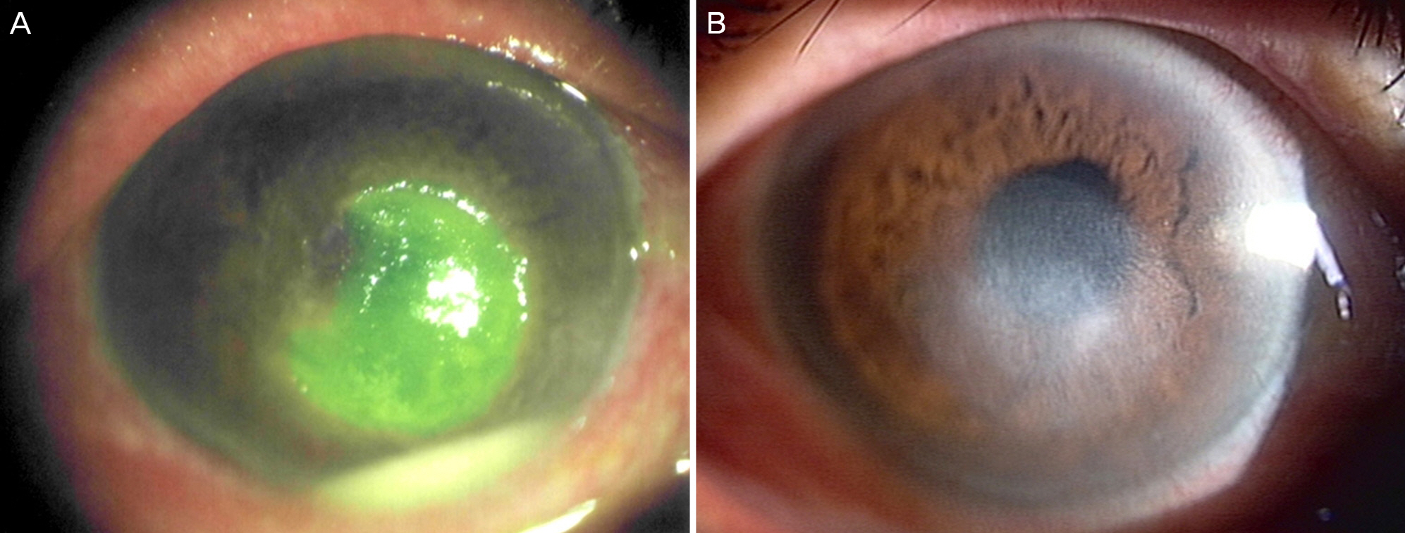
Figure 1. Slit-lamp photographs of Case 1. (A) At the initial visit, 4.5 × 4.5 mm sized epithelial defect with a 1.2 mm height hypopyon. (B) After 3 months, corneal lesion regressed with opacity at corneal center.
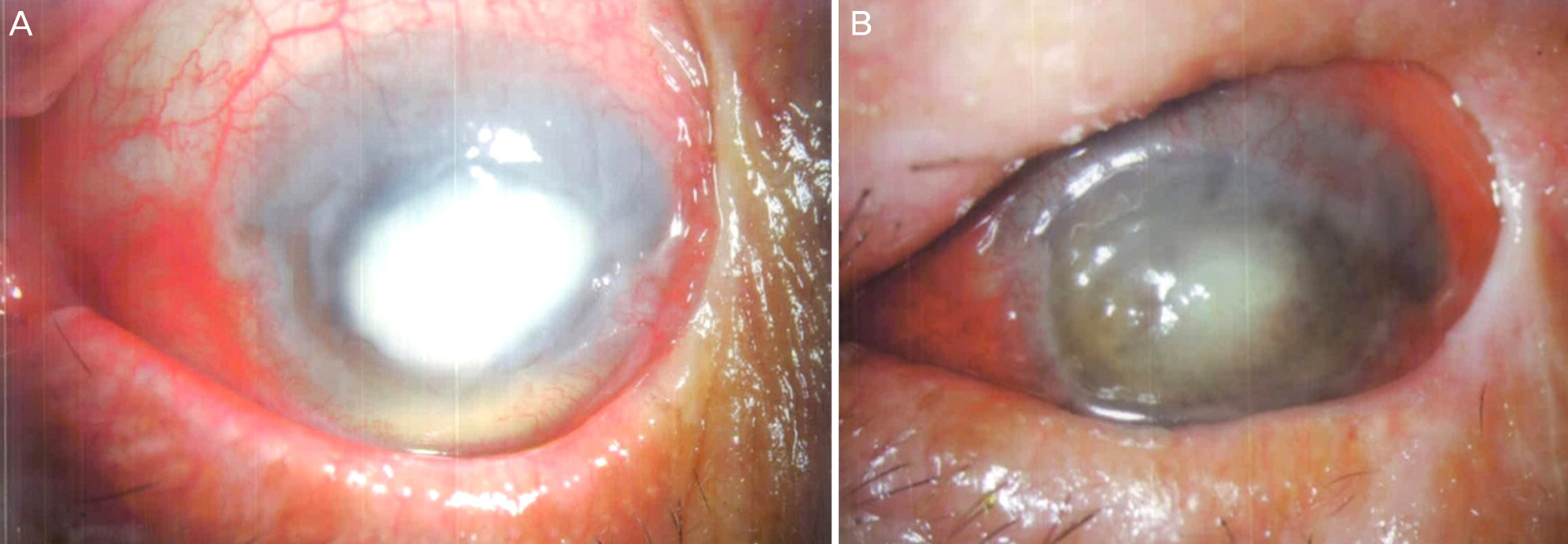
Figure 2. Slit-lamp photographs of Case 2. (A) At the initial visit, 6.0 × 7.0 mm sized epithelial defect with a 1.5 mm height hypopyon. (B) After 2 weeks, epithelial defect was healed with corneal opacity at the center and the infected eye became phthisis bulbi.

Figure 3. Slit-lamp photographs of Case 3. (A) At the initial visit, 8.0 × 7.0 mm sized epithelial defect with satellite lesion at the superior. (B) The patient rejected antifungal treatment and returned to hospital with corneal perforation.
Cited by 1 articles
-
Keratitis Caused by Paecilomyces lilacinus after Cataract Surgery in a Patient with Systemic and Autoimmune Disease
Shin Yeop Oh, Hye Sook Kang, Chang Kyu Lee
J Korean Ophthalmol Soc. 2016;57(11):1795-1800. doi: 10.3341/jkos.2016.57.11.1795.
Reference
-
References
1. Pastor FJ, Guarro J. Clinical manifestations, treatment and outcome of Paecilomyces lilacinus infections. Clin Microbiol Infect. 2006; 12:948–60.
Article2. Yuan X, Wilhelmus KR, Matoba AY, et al. Pathogenesis and outcome of Paecilomyces keratitis. Am J Ophthalmol. 2009; 147:691–6.e3.
Article3. Byun DS, Yang HN, Cho HG, Cha YJ. A case of corneal ulcer caused by Paecilomyces in diabetic patient wearing soft contact lens. J Korean Ophthalmol Soc. 1987; 28:667–71.4. Won JY, Shin JY, Hwang JH, Joo CK. A case of fungal keratitis caused by Paecilomyces lilacinus after penetrating keratoplasty. J Korean Ophthalmol Soc. 2014; 55:1384–7.5. Monden Y, Sugita M, Yamakawa R, Nishimura K. Clinical experience treating Paecilomyces lilacinus keratitis in four patients. Clin Ophthalmol. 2012; 6:949–53.
Article6. Hirst LW, Choong K, Playford EG. Nontraumatic paecilomyces anterior segment infection: a pathognomonic clinical appearance. Cornea. 2014; 33:1031–7.7. Yildiz EH, Ailani H, Hammersmith KM, et al. Alternaria and paecilomyces keratitis associated with soft contact lens wear. Cornea. 2010; 29:564–8.
Article8. Wu PC, Lai CH, Tan HY, et al. The successful medical treatment of a case of Paecilomyces lilacinus keratitis. Cornea. 2010; 29:357–8.
Article9. Arnoldner MA, Kheirkhah A, Jakobiec FA, et al. Successful treatment of Paecilomyces lilacinus keratitis with oral posaconazole. Cornea. 2014; 33:747–9.
Article10. McLintock CA, Lee GA, Atkinson G. Management of recurrent Paecilomyces lilacinus keratitis. Clin Exp Optom. 2013; 96:343–5.
Article11. Maier AK, Reichenbach A, Rieck P. Paecilomyces lilacinus keratitis. Ophthalmologe. 2011; 108:966–8.12. Yildiz EH, Abdalla YF, Elsahn AF, et al. Update on fungal keratitis from 1999 to 2008. Cornea. 2010; 29:1406–11.
Article13. Keay LJ, Gower EW, Iovieno A, et al. Clinical and microbiological characteristics of fungal keratitis in the United States, 2001-2007: a multicenter study. Ophthalmology. 2011; 118:920–6.
Article14. Lee KH, Chae HJ, Yoon KC. Analysis of risk factors for treatment failure in fungal keratitis. J Korean Ophthalmol Soc. 2008; 49:737–42.
Article15. Kim KH, Kim MJ, Tchah H. Management of fungal ocular infection with topical and intracameral voriconazole. J Korean Ophthalmol Soc. 2008; 49:1054–60.
Article16. Al-Badriyeh D, Neoh CF, Stewart K, Kong DC. Clinical utility of voriconazole eye drops in ophthalmic fungal keratitis. Clin Ophthalmol. 2010; 4:391–405.17. Sharma N, Sachdev R, Jhanji V, et al. Therapeutic keratoplasty for microbial keratitis. Curr Opin Ophthalmol. 2010; 21:293–300.
Article18. Cho SH, Park JW, Chung SK. The risk factor analysis of infectious corneal ulcers leading to eyeball removal. J Korean Ophthalmol Soc. 2008; 49:34–9.
Article
- Full Text Links
-
- Actions
-
Cited
- CITED
-
- Close
- Share
-
- Similar articles
-
- A Case of Fungal Keratitis Caused by Paecilomyces lilacinus after Penetrating Keratoplasty
- Recurrent Paecilomyces Keratitis in a Patient with Jones Tube after Conjunctivodacryocystorhinostomy
- Keratitis Caused by Paecilomyces lilacinus after Cataract Surgery in a Patient with Systemic and Autoimmune Disease
- Dendritic Keratitis Associated with Contact Lens Wear: a Case Series and Literature Review
- The Effect of Subconjunctival Injection of Tathion on Some Keratitis
