J Korean Ophthalmol Soc.
2010 Feb;51(2):248-253. 10.3341/jkos.2010.51.2.248.
Comparison of Retinal Nerve Fiber Layer Thickness in Early Normal-Tension Glaucoma and Early Primary Open-Angle Glaucoma
- Affiliations
-
- 1The Institute of Ophthalmology and Optometry, Department of Ophthalmology, Ewha Womans University School of Medicine, Seoul, Korea. ckrey02@ewha.ac.kr
- KMID: 2213147
- DOI: http://doi.org/10.3341/jkos.2010.51.2.248
Abstract
- PURPOSE
To investigate the comparison of retinal nerve fiber layer (RNFL) thickness parameters measured by optical coherence tomography (Stratus OCT 3000TM) and visual field indices in early normal-tension glaucoma (NTG) and early primary open-angle glaucoma (POAG).
METHODS
Sixty-one early normal-tension glaucomatous eyes, 21 early primary open-angle glaucomatous eyes and 34 healthy control eyes were enrolled in this cross-sectional study. Each subject received a visual field test (Humphrey C30-2) and the fast RNFL thickness algorithm test by OCT. The correlations between RNFL thickness and visual field indices were analyzed. The sensitivity and specificity for the detection of early glaucoma were determined with the area under the receiver operating characteristics curve (AUROC).
RESULTS
All RNFL thickness values except for the temporal quadrant RNFL thickness were significantly decreased in the early NTG and POAG groups (p<0.05). In early POAG, the average and superior quadrant RNFL thicknesses were significantly thinner than in the early NTG group. Significant correlations were observed between the PSD and the average and superior quadrant RNFL thicknesses in the early NTG and POAG groups (p<0.05). The average RNFL thickness for early glaucoma had the widest AUROC among all of the parameters.
CONCLUSIONS
In the early NTG group with visual field defects similar to those of early POAG, RNFL defects measured by OCT were less severe, particularly in the average and superior quadrant RNFLs.
Keyword
MeSH Terms
Figure
-
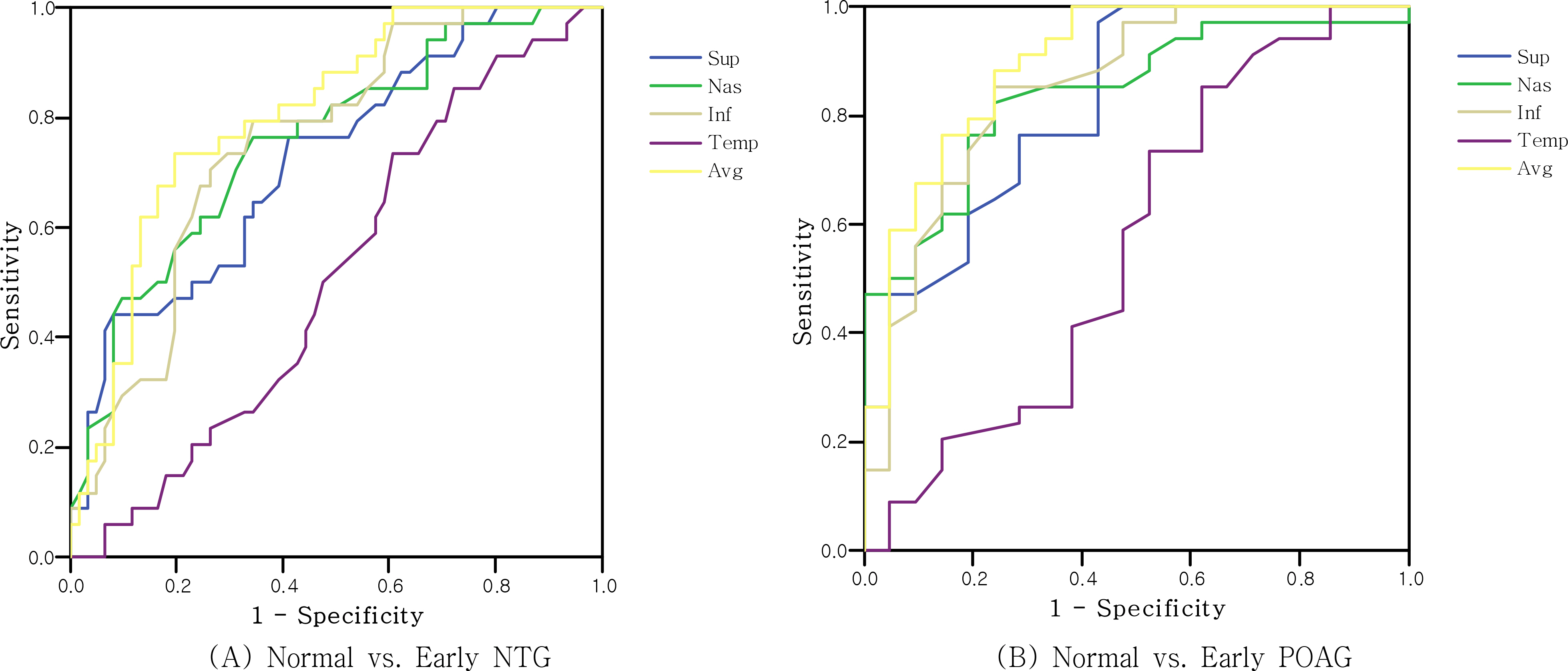
Figure 1. ROC curve for OCT parameters.
Reference
-
References
1. Quigley HA, Addicks EM, Green WR. Optic nerve damage in human glaucoma. III. Quantitative correlation of nerve fiber loss and visual field defect in glaucoma, ischemic neuropathy, aberrations, and toxic neuropathy. Arch Ophthalmol. 1982; 100:135–46.2. Sommer A, Katz J, Quigley HA, et al. Clinical detectable nerve fiber atrophy precedes the onset of glaucomatous field loss. Arch Ophthalmol. 1991; 109:77–83.3. Von Graefe A. ϋ ber die iridectomie bei glaucom und ϋ ber den aberrations usen prozess. Albercht von Graefes Arch Ophthalmol. 1857; 3:456–650.4. Levene RZ. Low-tension glaucoma: a critical review and new material. Surv Ophthalmol. 1980; 24:621–64.5. Kubota T, Khalil AK, Honda M, et al. Comparative study of retinal nerve fiber layer damage in Japanese patients with normal- and high-tension glaucoma. J Glaucoma. 1999; 8:363–6.
Article6. Kook MS, Sung K, Kim S, et al. Study of retinal nerve fiber layer thickness in eyes with high tension glaucoma and hemifield defect. Br J Ophthalmol. 2001; 85:1167–70.7. Yamazaki Y, Koide C, Miyazawa T, et al. Comparison of retinal nerve fiber layer in high- and normal-tension glaucoma. Graefes Arch Clin Exp Ophthalmol. 1991; 229:517–20.8. Woo SJ, Park KH, Kim DM. Comparison of localized nerve fiber layer defects in normal tension glaucoma and primary open angle glaucoma. Br J Ophthalmol. 2003; 87:695–8.9. Shields MB. Normal-tension glaucoma; is it different from aberrations open-angle glaucoma. Curr Opin Ophthalmol. 2008; 19:85–8.10. Mok KH, Lee VW, So KF. Retinal nerve fiber loss in high- and normal-tension glaucoma by optical coherence tomography. Optom Vis Sci. 2004; 81:369–72.
Article11. Quigley HA, Katz J, Derrick RJ, et al. An evaluation of optic disc and nerve fiber layer examinations in monitoring progression of early glaucoma damage. Ophthalmology. 1992; 99:19–28.
Article12. Shin IH, Kang SY, Kim CY, et al. Comparison of OCT and HRT finding among normal, normal tension glaucoma, and high aberrations glaucoma. Korean J Ophthalmol. 2008; 22:236–41.13. Kanamori A, Nakamura M, Escano MF, et al. Evaluation of the glaucomatous damage on retinal nerve fiber layer thickness aberrations by optical coherence tomography. Am J Ophthalmol. 2003; 135:513–20.14. Medeiros FA, Zangwill LM, Bowd C, et al. Evaluation of retinal nerve fiber layer, optic nerve head, and macular thickness aberrations for glaucoma detection using optical coherence aberrations. Am J Ophthalmol. 2005; 139:44–55.15. Park SE, Jung JK, Jung J-Y, Park SH. Optical coherence aberrations parameters of normal, glaucoma suspect, and early aberrations patients. J Korean Ophthalmol Soc. 2007; 48:1379–87.16. Leung CK, Mohamed S, Leung KS, et al. Retinal nerve fiber layer measurements in myopia: an optical coherence tomography study. Invest Ophthalmol Vis Sci. 2006; 47:5171–6.
Article
- Full Text Links
-
- Actions
-
Cited
- CITED
-
- Close
- Share
-
- Similar articles
-
- Clinical Evaluation of Unilateral Open-Angle Glaucoma: A Two-Year Follow-Up Study
- Comparison of the Central Retinal Vessel Diameter Between Glaucomatous and Normal Eye
- Choroidal Thickness in Primary Open-Angle Glaucoma Using Spectral-Domain Optical Coherence Tomography
- Influence of Diabetes Mellitus on the Retinal Ne rve Fiber Layer Thickness Measurement by Nerve Fiber Analyzer
- Ocular Dominance in Open-angle Glaucoma: The Shifting Trend Depending on Stage of the Disease
