J Korean Ophthalmol Soc.
2009 May;50(5):738-742. 10.3341/jkos.2009.50.5.738.
Comparison of the Central Retinal Vessel Diameter Between Glaucomatous and Normal Eye
- Affiliations
-
- 1Department of Ophthalmology, Korea University College of Medicine, Seoul, Korea. yongykim@mail.korea.ac.kr
- KMID: 2212312
- DOI: http://doi.org/10.3341/jkos.2009.50.5.738
Abstract
-
PURPOSE:To compare the diameter of central retinal vessels between patients with normal-tension glaucoma, primary open-angle glaucoma and healthy (control) eyes.
METHODS
The authors reviewed 30 eyes of 30 normal-tension glaucoma patients, 20 eyes of 20 primary open-angle glaucoma patients and 30 eyes of normal persons who had no systemic vascular diseases. The diameters of the central retinal arteries and veins were measured and calculated using a revised Parr-Hubbard formula, and results were compared between the groups.
RESULTS
The diameter of the central retinal vessel showed no statistical differences between the eyes with normal-tension glaucoma and primary-open angle glaucoma. However, there were significant differences between the eyes of patients with glaucoma and the normal control eyes (p<0.05). Conclusion: The diameter of the central retinal vessels in glaucoma patients were narrower than that in the control group. Our results suggest that the diameter of the central retinal vessels may affect the development of glaucoma both in normal tension and primary open-angle types, or that glaucomatous damage may influence the diameter of the central retinal vessels.
MeSH Terms
Figure
-
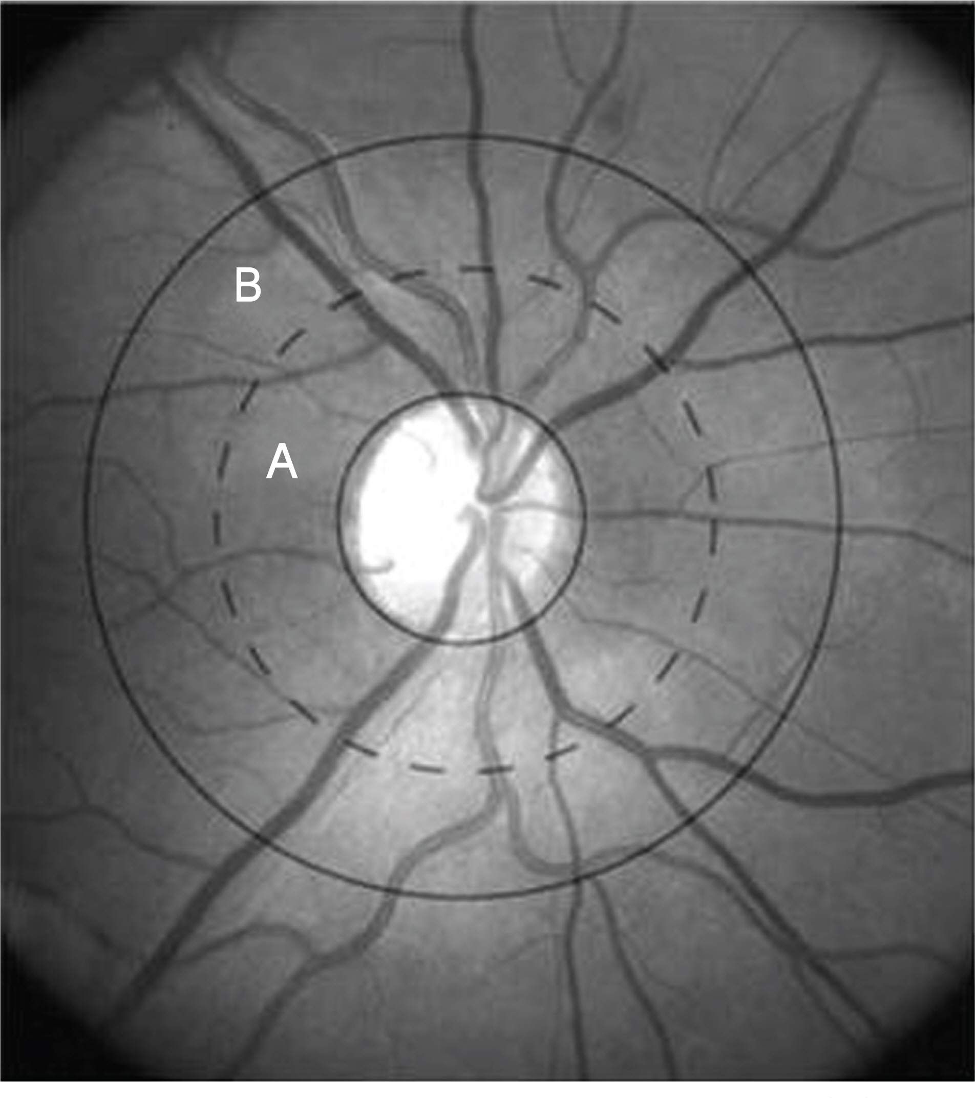
Figure 1. Digitized retinal photograph. Zone (A) is a half-disc diameter from the optic disc margin and Zone B is a half-disc to one and half-disc diameter from the optic disc margin. Retinal vessel diameter measurements are performed in Zone (B).

Figure 2. Comparison of the vessel diameters between 3 groups. Both CRAE and CRVE were significantly narrower in NTG and POAG group than innormal group. But there was no significant difference between NTG and POAG groups. *P<0.05 with multiple comparison test (Tukey B test).
Reference
-
References
1. Albert DM, Jokobiec FA. Principles and Practice of ophthalmology. Philadelphiia: WB Saunders Co.;1994. p. 1291.2. Shields MB. Textbook of Glaucoma. 2nd ed.Baltimore: Williams & Wilkins;1986. p. 145.3. Hollows FC, Graham PA. Intraocular preesure, glaucoma and glaucoma suspects in a defined population. Br J Ophthalmol. 1966; 50:570–86.4. Drance SM, Sweency VP, Morgan RW, Feldman F. Studies of factors involved in the production of low-tension glaucoma. Arch Ophthalmol. 1973; 89:457–85.5. Kahn HA, Leibowbitz HM, Ganley JP, et al. The Framigham eye study. I. Outline and major prevalence findings. Am J Epidemiol. 1977; 106:17–32.6. Hoskins HD, Kass MA. Becker-Shaffer's Diagnosis and therapy of the glaucoma. 6th ed.St. Louis: The CV Mosby Company;1989. p. 227.7. Drance SM, Douglas GR, Wijsman K. Response of blood flow to warm and cold in normal tension glaucoma patients. Am J Ophthalmol. 1988; 105:35–9.8. Mitchell P, Leung H, Wang JJ, et al. Retinal vessel diameter and open-andgle glaucoma. Ophthalmology. 2005; 112:245–50.9. Jonas JB, Nguyen XN, Anderson DR. Peripapillary retinal vessel diameter in normal and glaucoma eyes. I. Morphometric data. Invest Ophthalmol Vis Sci. 1989; 30:1599–603.10. Rader J, Feuer WJ, Anderson DR. Peripapillary vasoconstriction in the glaucomas and the anterior ischemic optic neuropathies. Am J Ophthalmol. 1994; 117:72–80.
Article11. Whelton PK, He J, Cutler JA, et al. Effects of oral potassium on blood pressure. Meta-analysis of randomized controlled clinical trials. JAMA. 1997; 277:1624–32.
Article12. Rankin SJ, Drance SM. Peripapillary focal retinal arteriolar narrowing in open angle glaucoma. J Glaucoma. 1996; 5:22–8.
Article13. Rankin SJ, Walman BE, Buckley AR, Drance SM. Color Doppler imaging and spectral analysis of the optic nerve vasculature in glaucoma. Am J Ophthalmol. 1995; 119:685–93.
Article14. Butt Z, O'Brien C, McKillop G, et al. Color Doppler imaging in untreated high- and normal-pressure open-angle glaucoma. Invest Ophthalmol Vis Sci. 1997; 38:690–6.15. Nicolela MT, Drance SM, Rankin SJ, et al. Color Doppler imaging in patients with asymmetric glaucoma and unilateral visual field loss. Am J Ophthalmol. 1996; 121:502–10.
Article16. Knudtson MD, Lee KE, Hubbard LD, et al. Revised formulars for summarizing retinal vessel diameters. Curr Eye Res. 2003; 27:143–9.17. Yamazaki Y, Hayamizu F. Comparison of flow velocity of ophthalmic artery between primary open angle glaucoma and normal tension glaucoma. Br J Ophthalmol. 1995; 79:732–4.
Article
- Full Text Links
-
- Actions
-
Cited
- CITED
-
- Close
- Share
-
- Similar articles
-
- Retinal Vessel Diameter: 1. Comparison of Normal and Glaucoma Eyes
- Retinal Vessel Diameter: 2. Its Correlation with Glaucomatous Optic Nerve Damage
- Non-glaucomatous Retinal Nerve Fiber Layer Defect Associated with Paravascular Inner Retinal Defect
- Analysis of Factors Associated with Retinal Vascular Caliber in Normal Korean Subjects
- Comparison between Glaucomatous and Non-glaucomatous Eyes with Unilateral Retinal Vein Occlusion in the Fellow Eye
