A Case of DiGeorge Syndrome With Ocular Manifestation
- Affiliations
-
- 1Department of Ophthalmology, Kyoungpook National University, College of Medicine, Daegu, Korea. jpshin@hitel.net
- 2Department of Ophthalmology, Pusan National University, College of Medicine, Busan, Korea.
- KMID: 2213008
- DOI: http://doi.org/10.3341/jkos.2009.50.12.1909
Abstract
- PURPOSE
DiGeorge syndrome (chromosome 22q11.2 deletion syndrome) is a syndrome of multiple congenital anomalies characterized by hypoplasia or aplasia of the thymus and parathyroid, cardiovascular malformation, immune deficiency, cleft palate, characteristic facial features, and hypocalcemia. Ocular findings of DiGeorge syndrome are posterior embryotoxon, retinal vascular tortuosity, strabismus, ptosis, amblyopia and tilted optic disc. The authors present a case of DiGeorge syndrome with ocular manifestation not reported previously in Korea. Case summary: A six-year old female diagnosed with DiGeorge syndrome was referred to the authors' department within the hospital. The chief complaint was blurring vision in both eyes. Best corrected visual acuity of the right eye was 0.5 and of the left eye was 0.63. Cycloplegic refraction revealed high hyperopia and astigmatism in both eyes (OD: +7.25 Dsph; -2.5 Dcyl axis 180degrees, OS: +6.25 Dsph; -3.75 Dcyl axis 180degrees). In addition, hypertelorism, ptosis and tortuous retinal vessels during fundus examination were noted.
CONCLUSIONS
Upon the initial diagnosis of DiGeorge syndrome in children, a comprehensive ocular examination is necessary because other ocular conditions may exist which can affect the visual development of the patient.
MeSH Terms
Figure
-
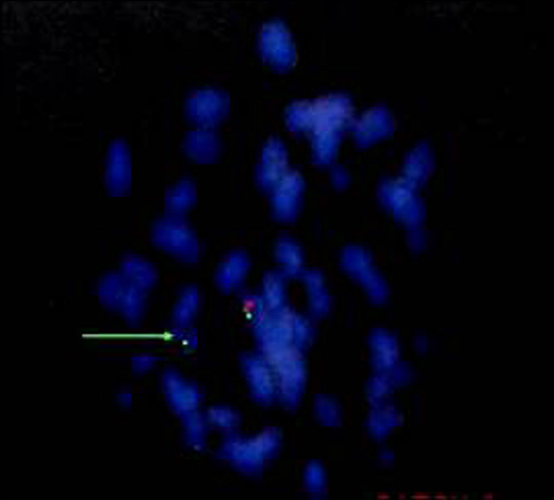
Figure 1. Result of fluorescence in situ hybridization (FISH) in this case. A submicroscopic deletion of chromosome 22q11.2 is detected by FISH with two labeled probes. The control probe (ARSA, 22q13.3, labeled green) binds to the end of chromosome 22, whereas the other probe (Tuple 1, 22q11.2, labeled red) binds to a sequence in the region that is commonly deleted in individuals with Di-George syndrome. The chromosome binds only with the ARSA probe, indicative of deletion of this region (green arrow).

Figure 2. Photographs of the patient. She had no characteristic facial feature of DiGoerge syndrome, but had ptosis of the left eye and hypertelorism.
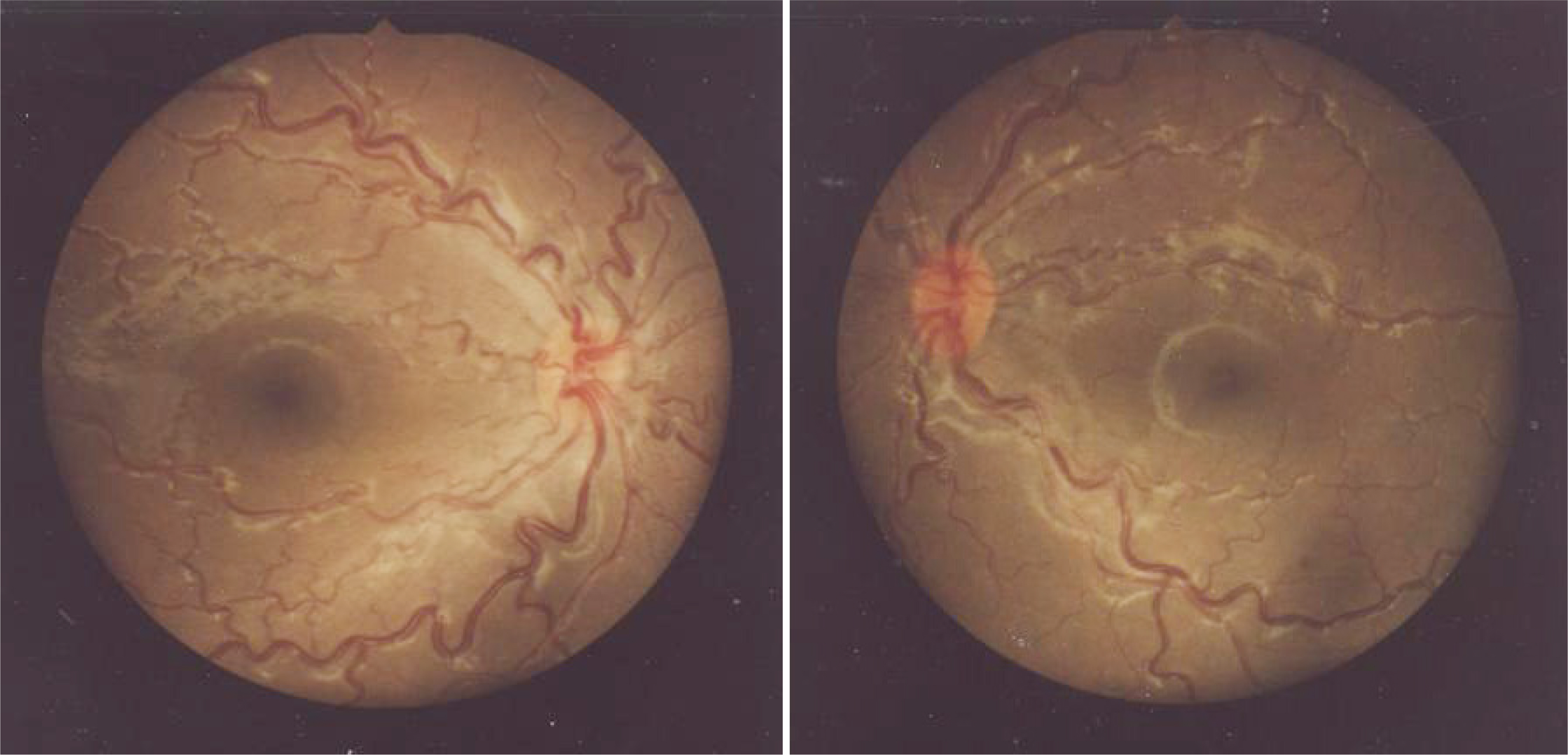
Figure 3. Fundus photographs of a case of Di-George syndrome. Retinal vascular tortuosity was noted in this case and this finding was known as relatively frequent ocular features in Di-George syndrome.
Reference
-
References
1. Forbes BJ, Binenbaum G, Edmond JC, et al. Ocular findings in the chromosome 22q11.2 deletion syndrome. J AAPOS. 2007; 11:179–82.
Article2. Kobrynski LJ, Sullivan KE. Velocardiofacial syndrome, Di-George syndrome: the chromosome 22q11.2 deletion syndromes. Lancet. 2007; 370:1443–52.
Article3. Sung TJ, Ko EY, Kim DH, et al. A Case of Partial Di-George Syndrome in Prematurity. J Korean Pediatr Soc. 2002; 45:383–9.4. Kim DJ, Hyun YY, Choi HM, et al. Di-George syndrome diagnosed by hypocalcemic tetany. Korean J Med. 2006; 70:299–301.5. Mansour AM, Goldberg RB, Wang FM, Shprintzen RJ. Ocular findings in the velocardiofacial syndrome. J Pediatr Ophthalmol Stra-bismus. 1987; 24:263–6.
Article6. Kok LL, Crewther SG, Crewther DP, Klistorner A. Visual function in velocardiofacial syndrome. Aust N Z J Ophthalmol. 1996; 24:53–5.
Article7. Binenbaum G, McDonald-McGinn DM, Zackai EH, et al. Sclero-cornea associated with the chromosome 22q11.2 deletion syndrome. Am J Med Genet A. 2008; 146:904–9.
Article8. Girgis RA, McKee HD, Innes JR. Swollen optic discs in a patient with the chromosome 22q11.2 deletion syndrome. Br J Ophtalmol. 2004; 88:591–2.
Article9. Hill VE, Pietucha S, Ells AL. Congenital vascular tortuosity in Di-George syndrome mimicking significant retinopathy of prematurity. Arch Ophthalmol. 2004; 122:132–3.
- Full Text Links
-
- Actions
-
Cited
- CITED
-
- Close
- Share
-
- Similar articles
-
- Various Psychiatric Manifestation in DiGeorge Syndrome (22q11.2 Deletion Syndrome): A Case Report
- Aspiration pneumonia in the child with DiGeorge syndrome: A case report
- A Case of Partial DiGeorge Syndrome
- Large Congenital Epigastric Hernia with DiGeorge Syndrome: A Case Report
- Complex cardiac Anomaly associated with the Digeorge syndrome
