J Korean Ophthalmol Soc.
2009 Jul;50(7):1115-1119. 10.3341/jkos.2009.50.7.1115.
Eight Cases of Descemet's Stripping Automated Endothelial Keratoplasty in Eyes With Bullous Keratopathy
- Affiliations
-
- 1Department of Ophthalmology and Visual Science, Seoul St. Mary's Hospital, College of Medicine, The Catholic University of Korea, Seoul, Korea. ckjoo@catholic.ac.kr
- KMID: 2212504
- DOI: http://doi.org/10.3341/jkos.2009.50.7.1115
Abstract
- PURPOSE
To investigate the postoperative results of Descemet's stripping automated endothelial keratoplasty in bullous keratoplasty and graft failure cases. CASE SUMMARY: Eight eyes of eight patients who underwent DSAEK between September 2006 and August 2008 were followed-up for at least 3 months, and the charts of eight patients were reviewed retrospectively. Five eyes of the total eight had bullous keratopathy, and three eyes needed DSAEK due to graft failure. The best corrective visual acuity (logMAR) improved from 1.625+/-0.60 to 0.825+/-0.35 and corneal thickness decreased from 785.63+/-135.58 microm to 692.00+/-150.85 microm 3 months postoperatively. Grafted corneal dislocation occurred in two eyes, and we repositioned the cornea in those participants by air injection. We found graft rejection signs in two cases, and one case showed graft failure despite steroid therapy. CONCLUSIONS: We think that DSAEK will be a good surgical procedure in bullous keratopathy or graft failure patients because of its favorable postoperative prognosis.
MeSH Terms
Figure
-
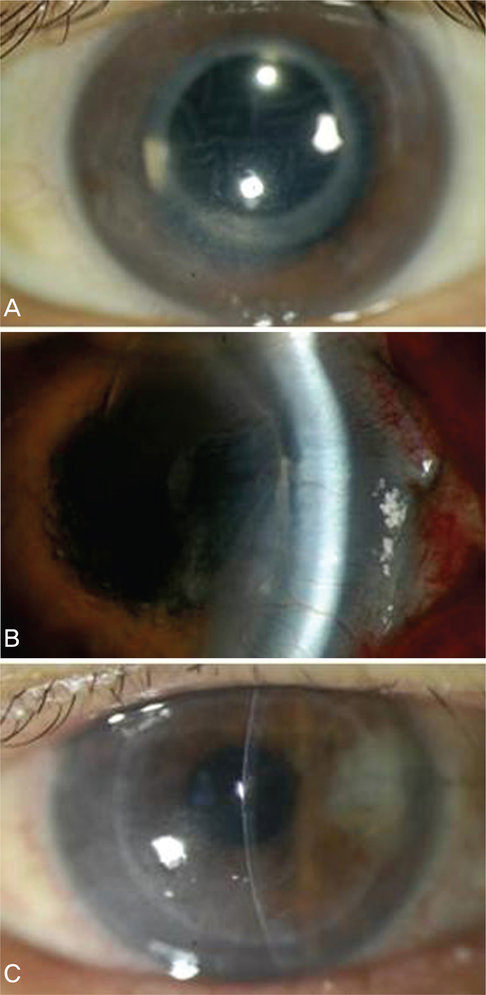
Figure 1. Preoperative and postoperative photographs (Case 3): Graft dislocation. (A) Preoperative picture. (B) Picture which shows graft dislocation (Postoperative 10 days). (C) Picture after air injection and graft reposition (Postoperative 1 month).

Figure 2. Preoperative and postoperative photographs (Case 2): Graft failure. (A) Preoperative picture. (B) Graft failure state (Postoperative 3 months).
Reference
-
References
1. Thompson RW Jr, Price MO, Bowers PJ, Price FW Jr. Long-term graft survival after penetrating keratoplasty. Ophthalmology. 2003; 110:1396–402.
Article2. Ing JJ, Ing HH, Nelson LR, et al. Ten-year postoperative results of penetrating keratoplasty. Ophthalmology. 1998; 105:1855–65.
Article3. Boisjoly HM, Tourigny R, Bazin R, et al. Risk factors of corneal graft failure. Ophthalmology. 1993; 100:1728–35.
Article4. Al-Towerki AE, Gonnah el-S, Al-Rajhi A, Wagoner MD. Changing indications for corneal transplantation at the King Khaled Eye Specialist Hospital (1983–2002). Cornea. 2004; 23:584–8.
Article5. Al-Yousuf N, Mavrikakis I, Mavrikakis E, Daya SM. Penetrating keratoplasty: indications over a 10 year period. Br J Ophthalmol. 2004; 88:998–1001.
Article6. Kang PC, Klintworth GK, Kim T, et al. Trends in the indications for penetrating keratoplasty (1980–2001). Cornea. 2005; 24:801–3.
Article7. Melles GR, Eggink FA, Lander F, et al. A surgical technique for posterior lamellar keratoplasty. Cornea. 1998; 17:618–26.
Article8. Terry MA, Ousley PJ. Deep lamellar endothelial keratoplasty in the first United States patients: early clinical results. Cornea. 2001; 20:239–43.9. Price FW Jr, Price MO. Descemet's stripping with endothelial keratoplasty in 50 eyes; a refractive neutral cornea transplant. J Refract Surg. 2005; 21:339–45.10. Gorovoy MS. Descemet-stripping automated endothelial keratoplastyaccommodative. Cornea. 2006; 25:886–9.11. Vincenzo S, Patricia T. Descemet-stripping automated endothelial keratoplasty by using suture for donor insertion. Cornea. 2008; 27:825–9.12. Koenig SB, Covert DJ. Early results of small incision Descemet's stripping and automated endothelial keratoplasty. Ophthalmology. 2007; 114:221–6.13. Koenig SB, Covert DJ, Dupps WJ Jr, Meisler DM. Visual acuity, refractive error, and endothelial cell density six months after DSAEK. Cornea. 2007; 26:670–4.14. Price MO, Price FW Jr. DSEK: comparative outcomes with micro-keratome-dissected and manually dissected donor tissue. Ophthalmology. 2006; 113:1936–42.15. Tan DT, Mehta JS. Future directions in lamellar corneal transplantation. Cornea. 2007; 26:S21–8.
Article16. Olson RJ. Air and the corneal endothelium: an in vivo specular microscopy study in cats. Arch Ophthalmol. 1980; 98:1283–4.17. Koenig GB, Covert DJ. Early results of small-incision Descemet's stripping and automated endothelial keratoplasty. Ophthalmology. 2006; 114:221–6.
Article18. Seo WM, Kim HK. Early result of Femtosecond laser assisted Descemet's membrane stripping endothelial keratoplasty. J Korean Ophthalmol Soc. 2008; 49:40–7.
Article19. Cheng YY, Pels E, Nuijts RM. Femtosecond laser assisted Descemet's membrane stripping endothelial keratoplasty. J Cataract Refract Surg. 2007; 33:152–5.20. Mehta JS, Por YM, Poh R, et al. Comparison of donor insertion techniques for DSAEK. Arch Ophthalmol. 2008; 126:1383–8.21. Busin M, Bhatt PR, Scorcia V. A modified technique for DSAEK to minimize endothelial cell loss. Arch Ophthalmol. 2008; 126:1133–7.22. Melles GR. Posterior lamellar keratoplasty: DLEK to DSEK to DMEK. Cornea. 2006; 25:879–81.
- Full Text Links
-
- Actions
-
Cited
- CITED
-
- Close
- Share
-
- Similar articles
-
- Early Result of Femtosecond Laser Assisted Descemet's Membrane Stripping Endothelial Keratoplasty
- Descemet Membrane Endothelial Keratoplasty to Treat Graft Failure after Descemet Stripping Endothelial Keratoplasty
- Factors Influencing Corneal Endothelial Cell Density after Descemet Stripping Automated Endothelial Keratoplasty
- Secondary Descemet Stripping Automated Endothelial Keratoplasty without Removing the Previously Grafted Posterior Stroma
- Comparison of Long-term Clinical Outcomes between Descemet's Stripping Automated Endothelial Keratoplasty and Penetrating Keratoplasty in Patients with Bullous Keratopathy
