More than 7-year survival of a patient following repeat hepatectomy for total 20 colon cancer liver metastases
- Affiliations
-
- 1Division of Hepato-Biliary-Pancreatic Surgery and Transplantation, Department of Surgery, Pusan National University Yangsan Hospital, Pusan National University School of Medicine, Yangsan, Korea. liversurgeon@hanmail.net
- 2Research Institute for Convergence of Biomedical Science and Technology, Pusan National University Yangsan Hospital, Yangsan, Korea.
- KMID: 2212213
- DOI: http://doi.org/10.4174/jkss.2012.82.2.128
Abstract
- A 54-year-old man was transferred with sigmoid colon cancer combined with multiple bilobar liver metastases. Nine metastases were in the left lobe and 5 metastases were in the right lobe. After low anterior resection, all 9 lesions in the left lobe were completely removed by wedge resections. Because the remnant liver volume after multiple wedge resection of the left lobe was not sufficient to perform a right hepatectomy simultaneously, we planned a two-stage hepatectomy. Right portal vein embolization was performed one week after the first liver operation. A right hepatectomy was safely performed 22 days after the first hepatectomy. A recurrent mass developed in the segment III 18 months after the right hepatectomy. Radiofrequency ablation (RFA) was performed to remove that lesion. Five other metastases developed 18 months after RFA whereby multiple wedge resections were performed. The patient has survived for more than 7 years after the first liver operation.
Keyword
MeSH Terms
Figure
-

Fig. 1 Initial abdominal ultrasonography and computed tomography. Four lesions in left lobe and 5 lesions in right lobe were found (white arrow, metastases in left lobe; black arrow, metastases in right lobe).

Fig. 2 (A) Abdominal computed tomography (CT) that was checked the day after first hepatectomy. Expected remnant liver volume was measured to be 29.5%. (B) Abdominal CT that was performed three weeks after first hepatectomy. Expected remnant liver volume increased to 35.8%. (C) Hypertrophied left lobe found in second hepatectomy. (D) Right hepatectomy was performed according to anatomical demarcation line.

Fig. 3 (A) Abdominal computed tomography (CT) taken at 15 months after second-stage hepatectomy showed no recurrent lesion. (B) Magnetic resonance image taken at 18 months after second-stage hepatectomy. Single recurrent mass (arrow) developed in remaining liver. (C) Abdominal CT checked after radiofrequency ablation (arrow, post-radiofrequency ablation lesion). (D) Abdominal CT taken at 14 months after radiofrequency ablation showed no other recurrent lesion (arrow, post-radiofrequency ablation lesion).
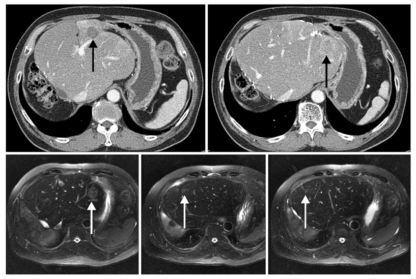
Fig. 4 Abdominal computed tomography and magnetic resonance image taken at 36 months after second-stage hepatectomy (arrows, multiple recurrent lesions).

Fig. 5 Abdominal computed tomography taken at 7 years after initial hepatectomy. No recurrent lesion was found.
Reference
-
1. Lochan R, White SA, Manas DM. Liver resection for colorectal liver metastasis. Surg Oncol. 2007. 16:33–45.2. Scheele J, Stang R, Altendorf-Hofmann A, Paul M. Resection of colorectal liver metastases. World J Surg. 1995. 19:59–71.3. Tanaka K, Shimada H, Ueda M, Matsuo K, Endo I, Togo S. Role of hepatectomy in treating multiple bilobar colorectal cancer metastases. Surgery. 2008. 143:259–270.4. Antoniou A, Lovegrove RE, Tilney HS, Heriot AG, John TG, Rees M, et al. Meta-analysis of clinical outcome after first and second liver resection for colorectal metastases. Surgery. 2007. 141:9–18.5. Minagawa M, Makuuchi M, Torzilli G, Takayama T, Kawasaki S, Kosuge T, et al. Extension of the frontiers of surgical indications in the treatment of liver metastases from colorectal cancer: long-term results. Ann Surg. 2000. 231:487–499.6. Imamura H, Sano K, Harihara Y, Noie T, Hasegawa K, Minagawa M, et al. Complete remission of disease for 5 years following initial and repeat resection of the liver for the removal of 22 metastases of colorectal origin. J Hepatobiliary Pancreat Surg. 2003. 10:321–324.7. Jaeck D, Bachellier P, Nakano H, Oussoultzoglou E, Weber JC, Wolf P, et al. One or two-stage hepatectomy combined with portal vein embolization for initially nonresectable colorectal liver metastases. Am J Surg. 2003. 185:221–229.8. Kokudo N, Tada K, Seki M, Ohta H, Azekura K, Ueno M, et al. Proliferative activity of intrahepatic colorectal metastases after preoperative hemihepatic portal vein embolization. Hepatology. 2001. 34:267–272.9. Adam R, Pascal G, Azoulay D, Tanaka K, Castaing D, Bismuth H. Liver resection for colorectal metastases: the third hepatectomy. Ann Surg. 2003. 238:871–883.10. Pawlik TM, Scoggins CR, Zorzi D, Abdalla EK, Andres A, Eng C, et al. Effect of surgical margin status on survival and site of recurrence after hepatic resection for colorectal metastases. Ann Surg. 2005. 241:715–722.
- Full Text Links
-
- Actions
-
Cited
- CITED
-
- Close
- Share
-
- Similar articles
-
- Repeat hepatectomy for recurred colorectal liver metastasis: is it justified?
- The Novel Approach of Multiple Colon Cancer Liver Metastases Treatment
- Clinical significance of post-hepatectomy hepatic failure in patients with liver metastases from colorectal cancer
- A Case of Hepatectomy for Liver Metastasis after Pancreatoduodenectomy for Carcinoma of Ampulla of Vater
- Application and Outcomes of Repeat Hepatectomy for Recurrent Hepatocellular Carcinom
