Hepatoid adenocarcinoma of the gallbladder with production of alpha-fetoprotein
- Affiliations
-
- 1Department of Surgery, Hanyang University College of Medicine, Seoul, Korea. hepafel@hanyang.ac.kr
- 2Department of Pathology, Hanyang University College of Medicine, Seoul, Korea.
- KMID: 2212191
- DOI: http://doi.org/10.4174/jkss.2011.80.6.440
Abstract
- Hepatoid adenocarcinoma (HAC) is a tumor with aberrant hepatocellular differentiation that occurs in extrahepatic organs. HAC of the gallbladder is rare, and cases of alpha-fetoprotein production are extremely rare. A 61-year-old man was diagnosed with gallbladder adenocarcinoma after laparoscopic cholecystectomy. A radical operation including resection of liver bed and lymph node dissection was performed, and no tumor cell was found. However, at postoperative 19 months, he showed lymphadenopathy of the portocaval area and tumor thrombi in the right portal vein with high levels of serum alpha-fetoprotein. After right hemihepatectomy and portahepatis lymph node dissection was performed, he was diagnosed with metastatic HAC. On reviewing the gallbladder specimen, the tumor finally demonstrated HAC as the primary origin. Despite adjuvant therapy, the patient died from multiple liver metastasis 26 months after cholecystectomy. Although HAC of the gallbladder is a very rare malignancy, awareness of its existence is critical to avoid misdiagnosis.
MeSH Terms
Figure
-
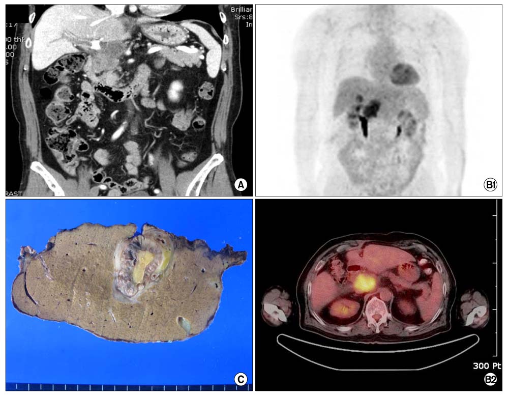
Fig. 1 (A) Abdominal computed tomography (CT) revealed the necrotic lymphadenopathy with infiltration of the portocaval area and thrombosis in the main and right portal vein. (B1 and B2) Positron emission tomography/CT showed the hypermetabolic lesion of 4 × 3 cm in portocaval area. (C) The cut surface of liver showed a well demarcated mass with a central bright yellowish area in the right portal vein.

Fig. 2 (A) Highly pleomorphic cells arranged in a rosettoid, nested, or trabecular pattern with relatively clear cytoplasm, enlarged nuclei, and prominent nucleoli. Numerous hyaline globules are noted in the intercellular or intracellular areas (H&E ×200). Immunohistochemical staining for alpha-fetoprotein (B) and alpha1-antitrypsin (C) showed strongly positive in the hepatoid area of the liver (×200).
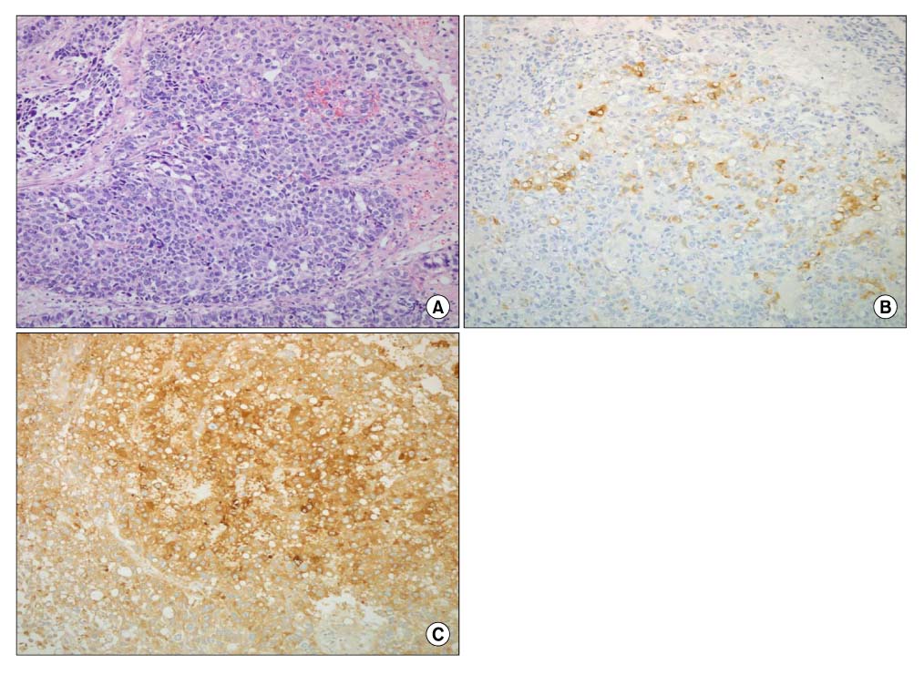
Fig. 3 (A) The gallbladder wall is replaced with infiltrating hepatoid adenocarcinoma composed of solid area, hepatoid carcinoma, with hyaline globules and conventional adenocarcinomatous area (H&E, ×200). Immunohistochemical staining for alpha-fetoprotein (B) and alpha1-antitrypsin (C) was positive in the hepatoid area of the previously operated gallbladder specimen (×200).
Reference
-
1. Supriatna Y, Kishimoto T, Uno T, Nagai Y, Ishikura H. Evidence for hepatocellular differentiation in alpha-fetoprotein-negative gastric adenocarcinoma with hepatoid morphology: a study with in situ hybridisation for albumin mRNA. Pathology. 2005. 37:211–215.2. Vardaman C, Albores-Saavedra J. Clear cell carcinomas of the gallbladder and extrahepatic bile ducts. Am J Surg Pathol. 1995. 19:91–99.3. Nakashima H, Nagafuchi K, Satoh H, Takeda K, Yamasaki T, Yonemasu H, et al. Hepatoid adenocarcinoma of the gallbladder. J Hepatobiliary Pancreat Surg. 2000. 7:226–230.4. Sakamoto K, Monobe Y, Kouno M, Moriya T, Sasano H. Hepatoid adenocarcinoma of the gallbladder: case report and review of the literature. Pathol Int. 2004. 54:52–56.5. Sakamoto K, Kimura N, Tokumura H, Ogasawara T, Moriya T, Sasano H. Hepatoid adenocarcinoma of the gallbladder. Histopathology. 2005. 47:649–651.6. Gakiopoulou H, Givalos N, Liapis G, Agrogiannis G, Patsouris E, Delladetsima I. Hepatoid adenocarcinoma of the gallbladder. Dig Dis Sci. 2007. 52:3358–3362.7. van den Bos IC, Hussain SM, Dwarkasing RS, Stoop H, Zondervan PE, Krestin GP, et al. Hepatoid adenocarcinoma of the gallbladder: a mimicker of hepatocellular carcinoma. Br J Radiol. 2007. 80:e317–e320.8. Ishikura H, Kishimoto T, Andachi H, Kakuta Y, Yoshiki T. Gastrointestinal hepatoid adenocarcinoma: venous permeation and mimicry of hepatocellular carcinoma, a report of four cases. Histopathology. 1997. 31:47–54.9. Maitra A, Murakata LA, Albores-Saavedra J. Immunoreactivity for hepatocyte paraffin 1 antibody in hepatoid adenocarcinomas of the gastrointestinal tract. Am J Clin Pathol. 2001. 115:689–694.10. Terracciano LM, Glatz K, Mhawech P, Vasei M, Lehmann FS, Vecchione R, et al. Hepatoid adenocarcinoma with liver metastasis mimicking hepatocellular carcinoma: an immunohistochemical and molecular study of eight cases. Am J Surg Pathol. 2003. 27:1302–1312.
- Full Text Links
-
- Actions
-
Cited
- CITED
-
- Close
- Share
-
- Similar articles
-
- Alpha-Fetoprotein-Producing Carcinoma of the Gallbladder: A case report
- A Case of Hepatoid Adenocarcinoma without Involving Liver Parenchyme
- Hepatoid Adenocarcinoma of the Stomach: A Case Report
- Hepatoid Adenocarcinoma of the Stomach: A Pathologic Analysis of 14 cases
- CT Findings of Hepatic Metastasis from Hepatoid Adenocarcinoma of the Rectum Mimicking Hepatocellular Carcinoma: A Case Report
