Upregulated Neuro-oncological Ventral Antigen 1 (NOVA1) Expression Is Specific to Mature and Immature T- and NK-Cell Lymphomas
- Affiliations
-
- 1Department of Pathology, Yonsei University College of Medicine, Seoul, Korea. soyoon@yuhs.ac
- 2Anatomic Pathology Reference Lab, Seegene Medical Foundation, Seoul, Korea.
- 3Department of Internal Medicine, Division of Hematology, Yonsei University College of Medicine, Seoul, Korea.
- KMID: 2211377
- DOI: http://doi.org/10.4132/jptm.2016.02.08
Abstract
- BACKGROUND
Recent studies have revealed that the splicing factor neuro-oncological ventral antigen 1 (NOVA1) is enriched in fibroblasts and accumulated T cells of tertiary lymphoid structures. In the present study, we investigated NOVA1 expression in various subtypes of mature and immature T- and natural killer (NK)-cell lymphomas as well as in various B-cell lymphoma subtypes.
METHODS
NOVA1 immunoexpression was evaluated in hyperplastic palatine tonsils (n = 20), T- and NK-cell lymphomas (n = 177), diffuse large B-cell lymphomas (n = 151), and other types of B cell lymphomas (n = 31). Nuclear staining intensity and percentage of positive tumor cells were graded. NOVA1 mRNA expression was analyzed in various lymphoma cell lines.
RESULTS
Tumor cells of T- and NK-cell lymphomas showed higher expression levels of NOVA1 than did normal paracortical T cells, and 56.5% of T- and NK-cell lymphoma cases showed diffuse and strong expression. The NOVA1 expression level varied according to the subtype; it was higher in angioimmunoblastic T-cell lymphoma, anaplastic lymphoma kinase (ALK)-negative anaplastic large cell lymphoma (ALCL), and T lymphoblastic leukemia/lymphoma (T-LBL), but it was lower in ALK-positive ALCL. In almost all B-cell lymphomas, NOVA1 expression was very low or negative. NOVA1 mRNA was also expressed in Jurkat, a T-LBL cell line.
CONCLUSIONS
The present findings suggest that NOVA1 upregulation may be involved in certain subtypes of T- and NK-cell lymphomas, but not in B-cell lymphomas. Upregulated NOVA1 expression seems to be a specific biological feature of activated T cells such as T- and NK-cell lymphomas.
Keyword
MeSH Terms
Figure
-
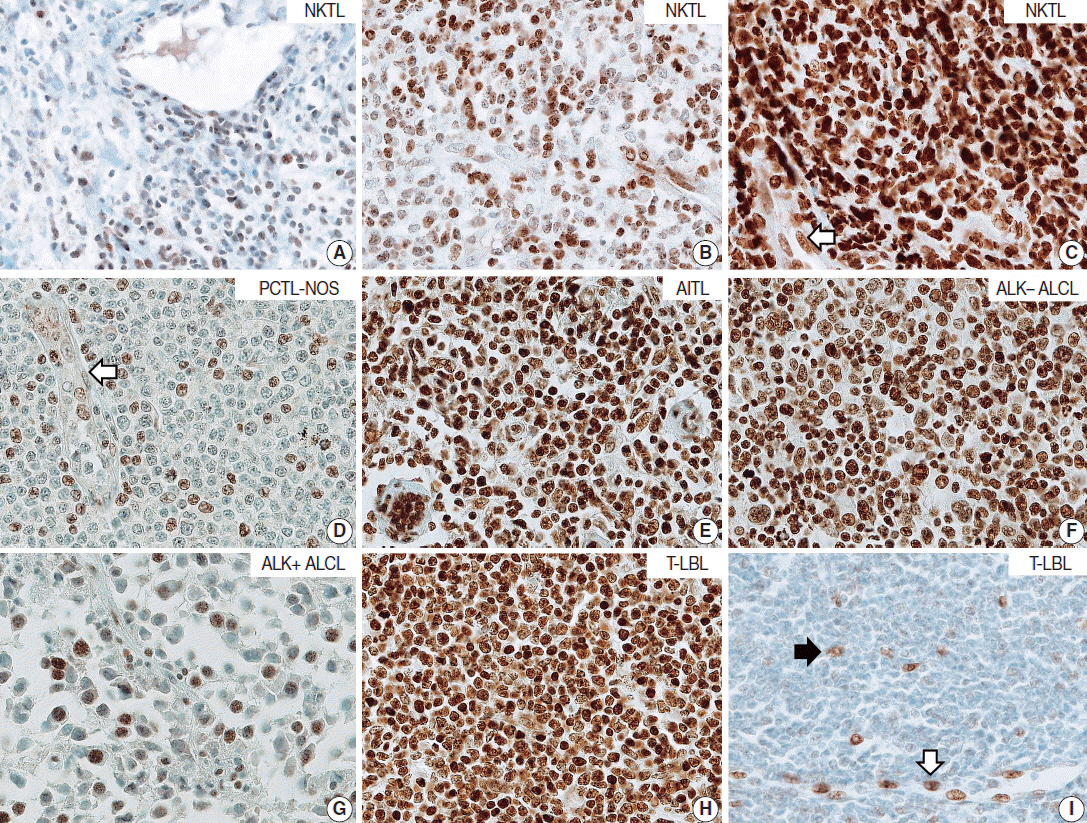
Fig. 1. Representative cases of T- and natural killer (NK)-cell lymphoma. Representative cases of extranodal NK/T-cell lymphoma, nasal type (NKTL) are presented, which show low (A), intermediate (B), and high (C) levels of neuro-oncological ventral antigen 1 (NOVA1) expression. Among mature (peripheral) T-cell lymphoma cases, low to high expression of NOVA1 is variably observed (D–G). (D) A case of peripheral T cell lymphoma, not otherwise specified (PTCL-NOS), reveals low NOVA1 expression. A case of angioimmunoblastic T-cell lymphoma (AITL) (E) and a case of anaplastic lymphoma kinase (ALK)-negative anaplastic large cell lymphoma (ALCL) (F) exhibits high NOVA1 expression. A case of ALK-positive ALCL (G) demonstrates low NOVA1 expression. A case of precursor T lymphoblastic lymphoma/leukemia (T-LBL) (H) shows high NOVA1 expression while a case of T-LBL shows low NOVA1 expression. Endothelial cells exhibit high or low NOVA1 expression in accordance with the NOVA1 status of tumor T cells (white arrow in panels in C and D). In cases where most tumor cells showed negative NOVA1 expression, stromal fibroblasts (black arrow in panel I) and endothelial cells (white arrow in panel I) demonstrate positive NOVA1 expression.

Fig. 2. Expression level of neuro-oncological ventral antigen 1 (NOVA1) in T- and natural killer (NK) cell lymphomas and normal T cells. (A) Expression level (score) of NOVA1 is higher in tumor cells of T/NK-cell lymphomas when compared to normal T cells of tonsil tissues (mean score, 7 vs 209; p < .001). (B) Within T/NK-cell lymphomas, T lymphoblastic leukemia/lymphoma (T-LBL) shows relatively higher levels of NOVA1 expression than do extranodal NK/T-cell lymphoma, nasal type (NKTL) or mature (peripheral) T-cell lymphomas, although a statistically significant difference is not noted (p = .226). (C) According to the specific subtypes of T/NK-cell lymphomas, the expression levels of tumor cells are different (p = .012). Angioimmunoblastic T-cell lymphoma (AITL), anaplastic lymphoma kinase (ALK)-negative anaplastic large cell lymphoma (ALCL), and T-LBL shows a relatively higher level of NOVA1 expression (arrow) when compared to other subtypes. ALK-positive ALCL reveals lower NOVA1 expression (dotted arrow) when compared to ALK-negative ALCL (mean score, 159 vs 256; p = .027) and ALK-positive ALCL shows the lowest expression among subtypes of T/NK cell lymphomas. (D) The rates of high, intermediate, and low expression of NOVA1 are different according to subtype (p < .001); high expression is relatively more frequent in AITL, ALK-negative ALCL, and T-LBL (solid arrow), while low expression is relatively more frequent in ALK-positive ALCL and NKTL (dotted arrow). The case number in the “others” category is too small (only 4 cases), and therefore, the NOVA1 expression may not be representative of this category. PTCL, peripheral T-cell lymphoma; PTCL-NOS, peripheral T-cell lymphoma, not otherwise specified.

Fig. 3. Neuro-oncological ventral antigen 1 (NOVA1) expression in B-cell lymphomas. (A, B) In the representative cases of diffuse large B-cell lymphoma (DLBCL), almost all tumor cells are negative for NOVA1. In some DLBCL cases, NOVA1-expressing fibroblasts/stromal support cells are occasionally found throughout the microenvironment (arrow in panel B). (C, D) In cases of mantle cell lymphoma (MCL) or follicular lymphoma (FL), tumor cells are negative for NOVA1, and fibroblasts/stromal support cells are positive for NOVA1 (arrow).
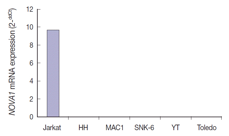
Fig. 4. Neuro-oncological ventral antigen 1 (NOVA1) mRNA expression in various cell lines. NOVA1 mRNA are expressed only in Jurkat cell lines, which correspond to T lymphoblastic leukemia/lymphoma. NOVA1 mRNA are not detected in other tested cell lines of HH, MAC1, SNK6, YT, or Toledo.
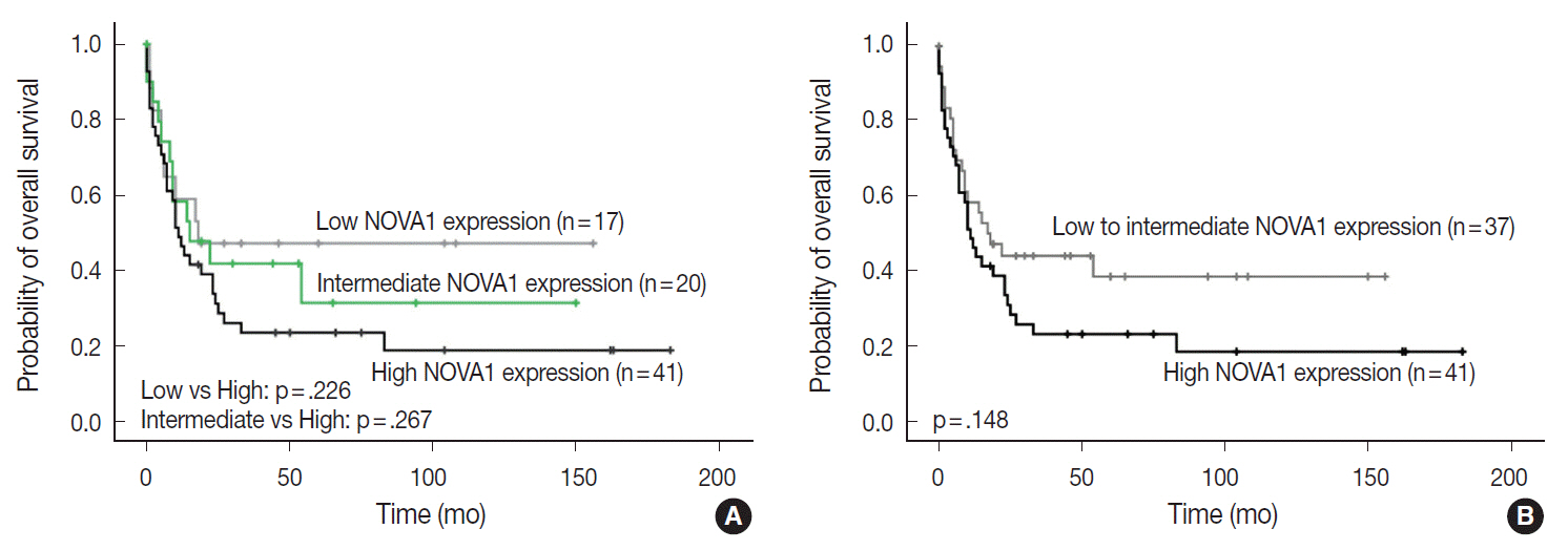
Fig. 5. Patient overall survival according to neuro-oncological ventral antigen 1 (NOVA1) expression in peripheral T-cell lymphoma, not otherwise specified; three-tiered (A) and two-tiered (B) analysis by Kaplan-Meier with log-rank test. High expression of NOVA1 tends to be related to shorter overall survival than low or intermediate expression, although statistical significance is not observed.
Reference
-
1. Barash Y, Calarco JA, Gao W, et al. Deciphering the splicing code. Nature. 2010; 465:53–9.
Article2. de la Grange P, Gratadou L, Delord M, Dutertre M, Auboeuf D. Splicing factor and exon profiling across human tissues. Nucleic Acids Res. 2010; 38:2825–38.
Article3. Merkin J, Russell C, Chen P, Burge CB. Evolutionary dynamics of gene and isoform regulation in mammalian tissues. Science. 2012; 338:1593–9.
Article4. Kalsotra A, Cooper TA. Functional consequences of developmentally regulated alternative splicing. Nat Rev Genet. 2011; 12:715–29.
Article5. Mallinjoud P, Villemin JP, Mortada H, et al. Endothelial, epithelial, and fibroblast cells exhibit specific splicing programs independently of their tissue of origin. Genome Res. 2014; 24:511–21.
Article6. Yoon SO, Kim EK, Lee M, et al. NOVA1 inhibition by miR-146b-5p in the remnant tissue microenvironment defines occult residual disease after gastric cancer removal. Oncotarget. 2016; 7:2475–95.
Article7. Drayton DL, Liao S, Mounzer RH, Ruddle NH. Lymphoid organ development: from ontogeny to neogenesis. Nat Immunol. 2006; 7:344–53.
Article8. Ruddle NH, Akirav EM. Secondary lymphoid organs: responding to genetic and environmental cues in ontogeny and the immune response. J Immunol. 2009; 183:2205–12.
Article9. Buckley CD, Barone F, Nayar S, Bénézech C, Caamaño J. Stromal cells in chronic inflammation and tertiary lymphoid organ formation. Annu Rev Immunol. 2015; 33:715–45.
Article10. Kalluri R, Zeisberg M. Fibroblasts in cancer. Nat Rev Cancer. 2006; 6:392–401.
Article11. Swerdlow SH, Campo E, Harris NL, et al. WHO classification of tumours of haematopoietic and lymphoid tissues. 4th ed. Lyon: IARC Press;2008.12. Park E, Park SY, Kim H, et al. Membranous insulin-like growth factor-1 receptor (IGF1R) expression is predictive of poor prognosis in patients with epidermal growth factor receptor (EGFR)-mutant lung adenocarcinoma. J Pathol Transl Med. 2015; 49:382–8.13. Falini B, Nicoletti I, Bolli N, et al. Translocations and mutations involving the nucleophosmin (NPM1) gene in lymphomas and leukemias. Haematologica. 2007; 92:519–32.
Article14. Kelemen O, Convertini P, Zhang Z, et al. Function of alternative splicing. Gene. 2013; 514:1–30.
Article15. Oberdoerffer S, Moita LF, Neems D, Freitas RP, Hacohen N, Rao A. Regulation of CD45 alternative splicing by heterogeneous ribonucleoprotein, hnRNPLL. Science. 2008; 321:686–91.
Article16. O’Rourke JP, Ness SA. Alternative RNA splicing produces multiple forms of c-Myb with unique transcriptional activities. Mol Cell Biol. 2008; 28:2091–101.
Article17. Henry C, Deschamps M, Rohrlich PS, et al. Identification of an alternative CD20 transcript variant in B-cell malignancies coding for a novel protein associated to rituximab resistance. Blood. 2010; 115:2420–9.
Article
- Full Text Links
-
- Actions
-
Cited
- CITED
-
- Close
- Share
-
- Similar articles
-
- A Case of Aggressive NK/T-cell Lymphoma/Leukemia with Cutaneous Involvement in Adolescence
- Immunoreactivity of CD99 in Non-Hodgkin's Lymphoma: Unexpected Frequent Expression in ALK-positive Anaplastic Large Cell Lymphoma
- A Case of Nasal CD56+ NK/T Cell Lymphoma Mimicking Cellulitis which Developed after Persistent Orbital Swelling
- Basic immunohistochemistry for lymphoma diagnosis
- Therapeutic Outcome of Epstein-Barr Virus Positive T/NK Cell Lymphoma in the Upper Aerodigestive Tract
